
Page 2259 - Williams Hematology ( PDFDrive )
P. 2259
2234 Part XII: Hemostasis and Thrombosis Chapter 131: The Antiphospholipid Syndrome 2235
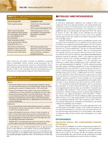
TABLE 131–1. Paths of Development of Antiphospholipid ETIOLOGY AND PATHOGENESIS
Assays: Historical Summary
Immunoassay Path Coagulation Path ETIOLOGY
As with most autoimmune conditions, the etiology of APS is not
1950s: Syphilis testing 1950s: Partial thromboplastin understood. It has been demonstrated that even normal healthy indi-
time inhibitor viduals have memory B cells that produce aPL antibodies. In a study
1970s: Lupus anticoagulant (LA) of patients with infectious mononucleosis, 10 to 60 percent of immu-
1980s: Antiphospholipid 1980s: Recognition that LAs noglobulin (Ig) M aPL-producing cells expressed CD27, the marker
(aPL) antibody enzyme-linked are inhibitors of phospholipid- of memory B cells. The affinity of aPL antibodies for their target
19
immunosorbent assay (ELISA; dependent coagulation becomes increased by the inclusion of amino acids lysine, arginine,
e.g., anticardiolipin [aCL] reactions and asparagine within the complementary determining regions of the
immunoassays) heavy and light chains. 20
1990s: Anticofactor ELISA Although antibodies against anionic phospholipid moieties arise
(anti-β -glycoprotein I [β GPI], during the course of infections such as syphilis and Lyme disease, those
2
2
antiprothrombin, etc.) are distinct from antibodies generated by patients with the syndrome
2005: Demonstration that 2004: Demonstration that because they generally recognize phospholipid epitopes directly (also
antibodies against domain I resistance to the anticoagulant referred to as “cofactor independent”) and are not associated with the
of β GPI are associated with effect of annexin A5 correlates clinical manifestations of the syndrome. There are intriguing hints
2
increased risk of thrombosis with thrombosis in antiphos- for molecular mimicry mechanisms and that infection and vaccina-
pholipid syndrome tion-induced APS could be related to autoimmune/autoinflammatory
syndrome induced by adjuvants (ASIA). aPL antibodies have been
21
reported in patients who developed thrombosis after varicella infec-
tion, 8,22,23 and in patients with hepatitis C. 24,25 aPL antibodies were
types of infections that induce formation of antibodies recognizing
anionic phospholipids directly, patients taking medications such as reported in a patient with cytomegalovirus (CMV) infection, mesen-
26,27
chlorpromazine or procainamide, and even in normal healthy individ- teric and femoropopliteal thrombosis. β GPI cofactor-dependent
2
uals. Testing of patients who have neither clinical manifestations of the antibodies against cardiolipin, phosphatidyl serine, and phosphatidyl
disorder or SLE for aPL antibodies should be discouraged because it ethanolamine have been identified in sera from patients with parvovi-
28
incurs the risk of inappropriate diagnostic and treatment decisions. rus B19. Bacterial infections are a predisposing risk factor for the cat-
astrophic form of APS (CAPS). A high proportion of HIV-1 patients
29
have aPL antibodies; more than 40 percent in one study, in which
18 percent had aCL and 30 percent had anti-β GPI (mostly of the IgA
2
TABLE 131–2. Sydney Investigational Criteria for Diagnosis isotype), ; however, positivity for these antibodies was not associated
30
of Antiphospholipid Syndrome with thrombosis. A link has been proposed between the cardiac valvu-
Clinical lar disease in acute rheumatic fever and the presence of aPL antibod-
31
• Vascular thrombosis (one or more episodes of arterial, venous, ies. aCL antibodies having β GPI dependence and LA activity have
2
or small vessel thrombosis). For histopathologic diagnosis, there been generated in rabbits immunized with lipid A and lipoteichoic
should not be evidence of inflammation in the vessel wall. acid, suggesting that some bacteria can contribute to the production
32
• Pregnancy morbidities attributable to placental insufficiency, of pathogenic aPL antibodies. It has also been proposed that cellu-
including three or more otherwise unexplained recurrent spon- lar apoptosis, with the resulting exposure of anionic phospholipids on
taneous miscarriages, before 10 weeks of gestation. Also, one cell surfaces, may trigger the generation of aPL antibodies. 33–35 Molec-
or more fetal losses after the 10th week of gestation, stillbirth, ular mimicry between β GPI-related synthetic peptides and structures
2
episode of preeclampsia, preterm labor, placental abruption, within bacteria, viruses, and tetanus toxoid have been demonstrated
36
intrauterine growth restriction or oligohydramnios that are oth- in an experimental model for APS. Mice immunized with a CMV-de-
37
erwise unexplained. rived peptide developed aPL antibodies and thrombosis, and showed
Laboratory evidence for endothelial cell activation. 38
39
• aCL or anti-β GPI IgG and/or IgM antibody present in medium Reports of familial clustering of raised aPL antibody levels indi-
2
or high titer on two or more occasions, at least 12 weeks apart, cate that genetic susceptibility can play a role in their development. In
measured by standard ELISAs. one study of 84 APS patients, more than 35 percent had at least one rela-
• Lupus anticoagulant in plasma, on two or more occasions, at tive, and more than 20 percent had two or more relatives, with evidence
least 12 weeks apart detected according to the guidelines of of at least one clinical feature of APS, such as thrombosis or recurrent
the International Society of Thrombosis and Hemostasis Scien- fetal loss. 40
tific Standardization Committee on Lupus Anticoagulants and
Phospholipid-Dependent Antibodies. PATHOGENESIS
• “Definite APS” is considered to be present if at least one of the Experimental Evidence That Antiphospholipid Antibodies
clinical criteria and one of the laboratory criteria are met.
Are Pathogenic
aCL, anticardiolipin; aPL, antiphospholipid; β GPI, β -glycoprotein I; It has been clearly established in a number of experimental animal mod-
2
2
ELISA, enzyme-linked immunosorbent assay; Ig, immunoglobulin. els for APS that aPL antibodies play a causal role in the development of
Data from Miyakis S, Lockshin MD, Atsumi T et al: International con- thrombosis and pregnancy loss. 41–45 Although it is reasonable to assume
sensus statement on an update of the classification criteria for defi- that the same holds for the human disorder, the epitopic specificities
nite antiphospholipid syndrome (APS). J Thromb Haemost 4:295–306, of the autoantibodies that cause disease and the mechanisms by which
206. they produce clinical manifestations require further elucidation.
Kaushansky_chapter 131_p2233-2252.indd 2234 9/18/15 5:10 PM