
Page 2262 - Williams Hematology ( PDFDrive )
P. 2262
2236 Part XII: Hemostasis and Thrombosis Chapter 131: The Antiphospholipid Syndrome 2237
98
TLRs, particularly TLR-2. This binding results in downstream signaling thrombosis. The presence of antibodies against EPCR in APS patients
that involves TRAF6 (tumor necrosis factor receptor-associated factor was proposed to be a risk factor for fetal death. 125
99
6) and MyD88 (myeloid differentiation factor 88). Increased expres- Antiphospholipid Antibodies Activate Platelets An experi-
sion of tissue factor is mediated by p38 mitogen-activated protein kinase mental animal model that includes in vivo imaging has provided data
(MAPK). 100 indicating that aPL-induced thrombosis is a consequence of platelet
Binding of autoantibodies to annexin A2 may also promote activation that then promotes endothelial activation and fibrin forma-
126
127
thrombosis by inhibiting fibrinolysis. APS patients have increased titers tion. aPL antibodies can induce platelet aggregation, an effect that
of antibodies against annexin A2, an endothelial surface receptor for might be promoted via signaling through apoER2 receptors; the β GPI
2
101
128
t-PA, and plasminogen. The blocking of annexin A2 by aPL antibodies binding site for apoER2 on platelets was localized to its domain V. As
impedes plasmin generation in a t-PA–dependent generation assay, and described above (see “Antigenic Specificities”), β GPI also has a damp-
2
inhibits cell surface plasmin generation on human umbilical vein endo- ening effect on platelet adhesion by interfering with the platelet–VWF
94
thelial cells. Several additional mechanisms have been identified by interaction, and consequently aPL antibodies, by interfering with this
which aPL antibodies can interfere with fibrinolysis. β GPI is a cofac- β GPI-mediated dampening, can increase platelet adhesion in flow
2
2
tor for t-PA–mediated activation of plasminogen, and aPL antibodies systems. 60
against β GPI interfere with its binding to t-PA, thereby downregulating Other Mechanisms APS patients have been shown to have
2
129
61
plasminogen activation. Finally, fibrinolysis may also be impaired by autoantibodies against tissue factor pathway inhibitor. Some aPL
autoantibodies directed against the catalytic site of plasmin or t-PA, 102,103 antibodies cross-react with heparin and heparinoid molecules, which
by an increased level of plasminogen activator inhibitor-1, and by are highly polyanionic, and hence, inhibit their contribution to antith-
104
69
inhibition of autoactivation of factor XII with ensuing reductions of rombin activity. aPL antibodies show cross-reactivity against oxidized
kallikrein and urokinase. 105 LDL and are associated with an increased risk of atherosclerosis.
131
130
Apolipoprotein E receptor 2 (apoER2), a member of the LDL- Antibodies against β GPI-oxidized LDL complexes have been proposed
2
132
106
receptor family is found on endothelial surfaces, monocytes, and to be atherogenic by reducing their clearance. Finally, in addition to
107
platelets, and may also serve as receptor for anti-β GPI–β GPI complexes promoting thrombosis, aPL antibodies may contribute to other vascu-
2
2
where it can also trigger the phosphatidylinositol 3′-kinase (PI3K)/Akt lar lesions by stimulating the mammalian target of rapamycin complex
108
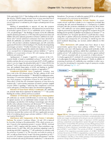
pathway and increase tissue factor and cell adhesion molecule expres- (mTORC) pathway (Fig. 131–4). 133
sion. IgG-mediated dimerization of β GPI and binding to ApoER2′
2
increases the sensitivity of platelets to agonists of aggregation. 109
Complement-Mediated Injury Complement activation may Thrombosis Antiphospholipid antibodies
play a role in the APS disease process. The IgG subtype of aPL most
2
closely correlates with thrombosis. 110,111 Blockade of complement activa-
tion using a C3 convertase inhibitor or genetic deletion of C3 protected Growth-factor Vasculopathy
receptors
Cytokine
mice from pregnancy complications induced by aPL antibodies. 112–114 receptors Integrins
These effects involve the aPL-stimulated expression of tissue factor by Cell
myeloid cells, and to involve proteinase-activated G-protein–coupled membrane
115
receptor (PAR)-2 signaling, indicating that complement activation
116
can be pathogenic via both direct injury and downstream signaling. Endothelial
Induction of Tissue Factor Activity in Leukocytes aPL antibodies P13K cell
can promote tissue factor expression by leukocytes. 115,117–119 The specific
binding site on these cells has not been elucidated. AKT mTORC2
Inhibition of Fibrinolysis and Endogenous Anticoagulation RICTOR
aPL antibodies can interfere with fibrinolysis in several ways. Antibod-
ies against annexin A2, an endothelial surface receptor for t-PA and mTORC1
plasminogen, can interfere with binding of plasminogen and t-PA and RAPTOR
thereby reduce plasmin formation and fibrinolysis. 94,101,103 Monoclonal
aPL antibodies derived from APS patients can directly inhibit plasmin’s Sirolimus
enzymatic activity. β GPI is a cofactor for t-PA–mediated activation
102
2
of plasminogen, and anti-β GPI can interfere with this activity. Also, Cell Cell Cell
120
2
it has been reported that women with APS have significantly increased growth proliferation survival
levels of circulating plasminogen activator inhibitor-1 (PAI-1), imply- Figure 131–4. Pathogenesis of vasculopathy in the antiphospholipid
ing impaired fibrinolysis. 104 syndrome. In addition to promoting thrombosis, antiphospholipid anti-
Interference with Components of the Protein C Activation bodies also trigger vasculopathy by binding to vascular endothelial cells
Pathway The protein C pathway (Chaps. 114 and 139) is initiated and activating the mammalian target of rapamycin (mTOR) signaling
by thrombin binding to thrombomodulin (TM), which activates pro- pathway. Extracellular and intracellular signals through the phosphati-
tein C bound to the endothelial protein C receptor (EPCR). Activated dylinositide 3′-kinase (PI3K)-AKT pathway activate mTOR pathway which
protein C (APC), together with free protein S, then proteolyses coag- regulates cell growth, proliferation and survival. The mTOR enzyme is a
ulation factors Va and VIIIa. APC also modulates signaling events by component of two complexes, mammalian target of rapamycin com-
plex (mTORC) 1 and mTORC2. The activity of mTORC1 is regulated by
interfering with PAR-1 signalling. 121,122 aPL antibodies can interfere with a subunit of the regulatory-associated protein of mTORC1 (RAPTOR),
the activation of protein C by TM–thrombin and with the activity of whereas the activity of mTORC2 is regulated by a subunit of the rapamy-
APC, as well as protect factors Va and VIIIa from proteolysis by APC. cin-insensitive companion of mTOR (RICTOR). (Modifed with permission
65
Acquired APC resistance has been described in APS plasmas and has from Eikelbloom JW, Weitz JI: The mTORC pathway in the antiphospholipid
123
124
been correlated with anti-β GPI domain I antibodies, a risk factor for syndrome. N Engl J Med 371(4):369–371, 2014.)
2
Kaushansky_chapter 131_p2233-2252.indd 2237 9/18/15 5:10 PM