
Page 2260 - Williams Hematology ( PDFDrive )
P. 2260
2234 Part XII: Hemostasis and Thrombosis Chapter 131: The Antiphospholipid Syndrome 2235
58
Antigenic Specificities lipoprotein (LDL) and may play a role in its clearance. β GPI binds to
2
Antibodies against phospholipid that arise during the immunologic lipopolysaccharide and the scavenged complex is taken up by mono-
59
response to syphilis and other infections (with the notable exception of cytes/macrophages. β GPI reduces platelet adhesion to collagen in
2
leprosy ) recognize anionic phospholipid epitopes directly, whereas flow chambers by interfering with the platelet–von Willebrand factor
46
47
pathogenic aPL antibodies recognize phospholipid-binding proteins, (VWF) interaction by binding to its A2 domain, thereby interfering
60
primarily β GPI. 48,49 with its binding to the platelet glycoprotein Ib complex. β GPI may
2
2
β GPI (also named apolipoprotein H), a member of the comple- also promote fibrinolysis as a cofactor for tissue-type plasminogen acti-
2
61
ment control protein or short consensus repeat superfamily, is a highly vator (t-PA) via its SCR domain V, which increases fibrinolytic activity.
50
glycosylated single-chain plasma protein composed of 326 amino The protein may have a further effect on fibrinolysis by binding to endo-
acids, with a molecular weight of approximately 50 kDa (Fig. 131–1). thelial cells via annexin A2, a protein that also serves as a receptor for
62
β GPI has five short consensus repeat (SCR) stretches of approximately plasminogen and t-PA. Homozygous β GPI-null mice have not been
2
2
63
60 amino acids (also referred to as complement control protein [CCP] demonstrated to display a thrombotic phenotype. However, the pro-
45
repeats). Epitopic specificities for individual domains may have patho- tein may play a role—though not a critical one—in the reproductive
genic and prognostic significance (see “Immunoassays” below). 51–54 process, as there was a reduction in the number of viable implantation
The affinity of β GPI for anionic phospholipids derives from cat- sites in β GPI-null mice and reduced fetal weight and fetal-to-placen-
2
2
ionic residues from its aminoterminus that have affinity for anionic tal weight ratio in late gestation, suggesting compromised placental
polar heads of phospholipids and a hydrophobic loop which inserts into function. 64
the lipid bilayer. β GPI has five domains for which antibodies have been In addition to β GPI, a number of other antigenic targets have been
2
2
identified. IgG antibodies against an epitope comprising Gly40-Arg43 identified for aPL, including, but not limited to, prothrombin, coagula-
in the domain I of β GPI have been reported to have a stronger correla- tion factor V, protein C, protein S, annexin A2, annexin A5, high- and
2
tion with thrombosis than antibodies against other epitopes. Recent low-molecular-weight kininogens, and factors VII/VIIa and vimentin–
51
data support the concept that β GPI undergoes conformational changes cardiolipin complex. 65–68
2
that may be important for the APS disease process. By transmission
electron microscopy, unbound β GPI appears to be in a closed confor-
2
mation because of the affinity of a portion of carboxyterminal domain Proposed Pathogenic Mechanisms
V for the protein’s amninoterminal domain I, where the phospholipid Table 131–3 and Fig. 131–2 summarize several of the main current
binding site is located near the carboxy-terminus of SCR domain hypotheses for pathogenic mechanisms in APS. The mechanisms of the
V (see Fig. 131–1). The binding of β GPI to anionic phospholipid human APS disease process have been difficult to elucidation, mainly
55
2
membranes, requires a conformational change which exposes an for two reasons: (1) The phenotypes of vascular thrombosis and preg-
epitope in domain I that had been cryptic in the unbound conformation nancy morbidity are not unique to APS, so it is difficult to ascertain
(see Fig. 131–1). 55 whether the candidate mechanism is playing a causal role or is incidental.
Although β GPI bind to phospholipids, its role in aPL-mediated (2) Antibodies isolated from APS patients recognize a multiplicity of
2
cell signaling (described in the section Proposed Pathogenic Mecha- antigenic determinants 69,70 that can have a broad range of effects, so it is
nisms), is mediated via binding to toll-like receptors (TLRs) and not by difficult to determine which specificities and effects are responsible for
direct binding to the lipid bilayer. disease manifestations in humans.
While the in vivo biologic function(s) of the protein has (have)
not been defined, several interesting properties have been demon-
strated. The molecule binds to apoptotic cells, and may play a role in TABLE 131–3. Proposed Pathogenic Mechanisms for
56
their phagocytosis and clearance. β GPI binds to oxidized low-density
57
2 Antiphospholipid Syndrome
I. Disruption of endothelial surface and annexin A5 anticoagu-
Arg 39 & 34 lant shield
DV DI DI
+ + + + Lys 19 II. Enhanced cell signaling
Binding site A. Mediated by antibodies against annexin A2
DIV DII DII of antibodies B. Mediated by antibodies to ApoE2R
DIII DIII C. Induction of endothelial surface proadhesive molecules
D. Induction of tissue factor expression on monocytes and
endothelial cells
DIV
E. Complement-mediated signaling and injury
DV III. Impeding of fibrinolysis and endogenous anticoagulation
+ + Lys 305 & 317
A. Interference with plasminogen and tissue plasminogen
activator
B. Interference with components of the protein C activation
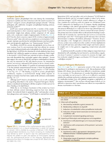
Figure 131–1. Schematic of the conformational states of β GPI. The pathway
2
unbound protein is in a closed conformation in which the epitope IV. Activation of platelets
on domain I (DI) is shielded by a portion of domain V (DV). Binding
to phospholipid membranes via a “barb,” consisting of a hydrophobic A. Interference with β -glycoprotein I dampening of von
2
loop formed by Ser311 to Lys317, near the carboxyterminus of DV, Willebrand factor–mediated platelet adhesion
requires the protein to open and exposes an immunogenic epitope V. Other mechanisms
near the aminoterminal portion of the molecule. (Reproduced with A. Mammalian target of rapamycin complex pathway–
permission from Rand JH A snappy new concept for APS. Blood 2010 Aug mediated vasculopathy
26;116(8):1193–1194.)
Kaushansky_chapter 131_p2233-2252.indd 2235 9/18/15 5:10 PM