
Page 232 - Williams Hematology ( PDFDrive )
P. 232
206 Part IV: Molecular and Cellular Hematology Chapter 15: Apoptosis Mechanisms: Relevance to the Hematopoietic System 207
FLIP TNFR1
The c-FLIP proteins are another type of apoptosis suppressor that oper-
ates via directly binding to certain caspases and their upstream activator,
FADD. The c-FLIP gene of humans resides in a tandem gene cluster on
chromosome 2, which contains the genes encoding procaspases 8 and TRADD TRADD TRADD
10, suggestive of gene duplication events. Two isoforms of c-FLIP are
produced from a single gene, including the long form, which is highly TRAF2 Rip1 FADD Rip1
similar in overall sequence to procaspases 8 and 10, containing tandem
copies of DEDs, followed by a pseudocaspase domain that lacks enzy-
matic activity. The shorter isoform consists only of the DED domains, cIAP1-2
thus resembling analogous proteins encoded in the genomes of some
mammalian viruses. FLIP-S is exclusively antoptotic whereas FLIP-L IKK Caspase 8/10 Rip3
52
can be either pro- or antiapoptotic, depending on its levels of expression
relative to procaspases 8 and 10. In general, FLIP proteins form com- IκB Caspase 3/7 MLKL
53
plexes with procaspases 8 and 10, preventing their dimerization and
activation, as well as competing for binding to adapter protein FADD, NF-κB
which is required for caspase recruitment to DR complexes. 54,55 Thus,
in most circumstances, FLIP proteins create blockades in the extrinsic Cell Survival Apoptosis Necroptosis
pathway for apoptosis.
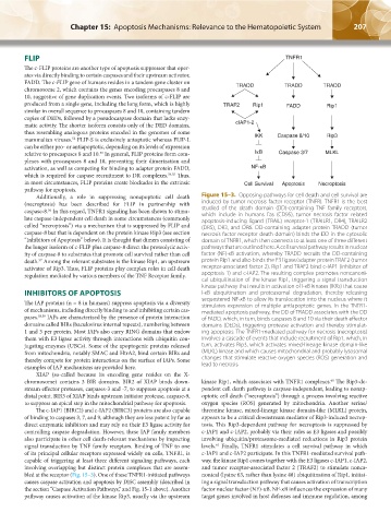
Additionally, a role in suppressing nonapoptotic cell death Figure 15–3. Opposing pathways for cell death and cell survival are
(necroptosis) has been described for FLIP in partnership with induced by tumor necrosis factor receptor (TNFR). TNFR1 is the best
caspase-8. In this regard, TNFR1 signaling has been shown to stimu- studied of the death domain (DD)-containing TNF family receptors,
56
which include in humans Fas (CD95), tumor necrosis factor–related
late caspase- independent cell death in some circumstances (commonly apoptosis-inducing ligand (TRAIL) receptor-1 (TRAILR1, DR4), TRAILR2
called “necroptosis”) via a mechanism that is suppressed by FLIP and (DR5), DR3, and DR6. DD-containing adapter protein TRADD (tumor
caspase-8 but that is dependent on the protein kinase Rip3 (see section necrosis factor receptor death domain) binds the DD in the cytosolic
“Inhibitors of Apoptosis” below). It is thought that dimers consisting of domain of TNFR1, which then connects to at least one of three different
the longer isoform of c-FLIP plus caspase-8 direct the proteolytic activ- pathways that are outlined here. A cell survival pathway results in nuclear
ity of caspase-8 to substrates that promote cell survival rather than cell factor (NF)-κB activation, whereby TRADD recruits the DD-containing
death. Among the relevant substrates is the kinase Rip1, an upstream protein Rip1 and also binds the E3 ligase/adapter protein TRAF2 (tumor
57
activator of Rip3. Thus, FLIP proteins play complex roles in cell death receptor- associated factor 2). Rip1 and TRAF2 bind c-IAP1 (inhibitor of
regulation mediated by various members of the TNF Receptor family. apoptosis 1) and c-IAP2. The resulting complex promotes noncanoni-
cal ubiquitination of the kinase Rip1, triggering a signal transduction
kinase pathway that results in activation of I-κB kinases (IKKs) that cause
INHIBITORS OF APOPTOSIS I-κB ubiquitination and proteasomal degradation, thereby releasing
The IAP proteins (n = 8 in humans) suppress apoptosis via a diversity sequestered NF-κB to allow its translocation into the nucleus where it
stimulates expression of multiple antiapoptotic genes. In the TNFR1-
of mechanisms, including directly binding to and inhibiting certain cas- mediated apoptosis pathway, the DD of TRADD associates with the DD
pases. 58,59 IAPs are characterized by the presence of protein interaction of FADD, which, in turn, binds caspases 8 and 10 via their death effector
domains called BIRs (baculovirus internal repeats), numbering between domains (DEDs), triggering protease activation and thereby stimulat-
1 and 3 per protein. Most IAPs also carry RING domains that endow ing apoptosis. The TNFR1-mediated pathway for necrosis (necroptosis)
them with E3 ligase activity through interactions with ubiquitin con- involves a cascade of events that include recruitment of Rip1, which, in
jugating enzymes (UBCs). Some of the apoptogenic proteins released turn, activates Rip3, which activates mixed-lineage kinase domain-like
from mitochondria, notably SMAC and HtrA2, bind certain BIRs and (MLKL) kinase and which causes mitochondrial and probably lysosomal
thereby compete for protein interactions on the surface of IAPs. Some changes that stimulate reactive oxygen species (ROS) generation and
examples of IAP mechanisms are provided here. lead to necrosis.
XIAP (so-called because its encoding gene resides on the X-
chromosome) contains 3 BIR domains. BIR2 of XIAP binds down- kinase Rip1, which associates with TNFR1 complexes. The Rip3-de-
60
stream effector proteases, caspases-3 and -7, to suppress apoptosis at a pendent cell death pathway is caspase-independent, leading to nonap-
distal point. BIR3 of XIAP binds upstream initiator protease, caspase-9, optotic cell death (“necroptosis”) through a process involving reactive
to suppress an apical step in the mitochondrial pathway for apoptosis. oxygen species (ROS) generated by mitochondria. Another serine/
The c-IAP1 (BIRC2) and c-IAP2 (BIRC3) proteins are also capable threonine kinase, mixed-lineage kinase domain-like (MLKL) protein,
of binding to caspases 3, 7, and 9, although they are less potent by far as appears to be a critical downstream mediator of Rip3-induced necrop-
direct enzymatic inhibitors and may rely on their E3 ligase activity for tosis. This Rip3-dependent pathway for necroptosis is suppressed by
controlling caspase degradation. However, these IAP family members c-IAP1 and c-IAP2, probably via their roles as E3 ligases and possibly
also participate in other cell death-relevant mechanisms by impacting involving ubiquitin/proteasome-mediated reductions in Rip3 protein
61
signal transduction by TNF family receptors. Binding of TNF to one levels. Finally, TNFR1 stimulates a cell survival pathway in which
of its principal cellular receptors expressed widely on cells, TNFR1, is c-IAP1 and c-IAP2 participate. In this TNFR1-mediated survival path-
capable of triggering at least three different signaling pathways, each way, the kinase Rip1 comes together with the E3 ligases c-IAP1, c-IAP2,
involving overlapping but distinct protein complexes that are assem- and tumor receptor-associated factor 2 (TRAF2) to stimulate nonca-
bled at the receptor (Fig. 15–3). One of these TNFR1-initiated pathways nonical (lysine 63, rather than lysine 48) ubiquitination of Rip1, initiat-
causes caspase activation and apoptosis by DISC assembly (described in ing a signal transduction pathway that causes activation of transcription
the section “Caspase Activation Pathways,” and Fig. 15-1 above). Another factor nuclear factor (NF)-κB. NF-κB influences the expression of many
pathway causes activation of the kinase Rip3, usually via the upstream target genes involved in host defenses and immune regulation, among
Kaushansky_chapter 15_p0203-0212.indd 207 17/09/15 6:37 pm