
Page 2368 - Williams Hematology ( PDFDrive )
P. 2368
2342 Part XIII: Transfusion Medicine Chapter 136: Erythrocyte Antigens and Antibodies 2343
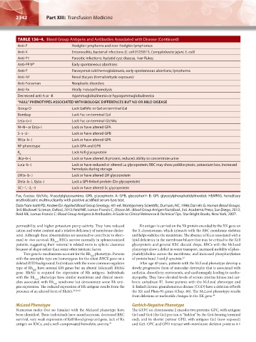
TABLE 136–4. Blood Group Antigens and Antibodies Associated with Disease (Continued)
Anti-I T Hodgkin lymphoma and non-Hodgkin lymphomas
Anti-K Enterocolitis, bacterial infections (E. coli 0125:B15, Campylobacter jejuni, E. coli)
Anti-P1 Parasitic infections: hydatid cyst disease, liver flukes
Anti-PP1P k Early spontaneous abortions
Anti-P Paroxysmal cold hemoglobinuria, early spontaneous abortions, lymphoma
Anti-NF Renal dialysis (formaldehyde exposure)
Anti-Forssman Neoplastic disorders
Anti-Rx Virally induced hemolysis
Decreased anti-A or -B Agammaglobulinemia or hypogammaglobulinemia
“NULL” PHENOTYPES ASSOCIATED WITH BIOLOGIC DIFFERENCES BUT NO OR MILD DISEASE
Group O Lack GalNAc or Gal on terminal Gal
Bombay Lack Fuc on terminal Gal
Le(a–b–) Lack Fuc on terminal GlcNAc
M–N– or En(a–) Lack or have altered GPA
S–s–U– Lack or have altered GPB
Wr(a–b–) Lack or have altered GPA
M phenotype Lack GPA and GPB
k
K Lack Kell glycoprotein
0
Jk(a–b–) Lack or have altered Jk protein, reduced ability to concentrate urine
Lu(a–b–) Lack or have reduced or altered Lu glycoprotein; RBC may show poikilocytosis, potassium loss, increased
hemolysis during storage
LW(a–b–) Lack or have altered LW glycoprotein
Do(a–b–), Gy(a–) Lack a GPI-linked protein (Do glycoprotein)
SC:–1,–2,–3 Lack or have altered Sc glycoprotein
Fuc, fucose; GlcNAc, N-acetylglucosamine; GPA, glycophorin A; GPB, glycophorin B; GPI, glycosylphosphatidylinositol; HEMPAS, hereditary
erythroblastic multinuclearity with positive acidified serum lysis test.
Data from Issitt PD, Anstee DJ: Applied Blood Group Serology, 4th ed. Montgomery Scientific, Durham, NC, 1998; Daniels G: Human Blood Groups,
3rd. Blackwell Science, Oxford, 2013; Reid ME, Lomas-Francis C, Olsson ML: Blood Group Antigen FactsBook, 3rd. Academic Press, San Diego, 2012;
Reid ME, Lomas-Francis C: Blood Group Antigens & Antibodies: A Guide to Clinical Relevance & Technical Tips. Star Bright Books, New York, 2007.
permeability, and higher potassium pump activity. They have reduced Kx antigen is carried on the Xk protein encoded by the XK gene on
cation and water content and a relative deficiency of membrane choles- the X chromosome, which interacts with the RBC membrane skeleton
terol. Although these abnormalities are assumed to contribute to short- and helps stabilize the membrane. The absence of Kx is associated with a
ened in vivo survival, Rh RBCs survive normally in splenectomized lipid deficiency in the membrane bilayer that may be critical to the Kell
null
patients, suggesting their removal is related more to splenic clearance glycoprotein and general RBC discoid shape. RBCs with the McLeod
because of shape rather than some other intrinsic factor. phenotype show a defect in water transport, increased mobility of phos-
Two genetic mechanisms account for the Rh phenotype. Persons phatidylcholine across the membrane, and increased phosphorylation
null
with the amorphic type are homozygous for the silent RHCE gene on a of protein band 3 and β-spectrin. 43
deleted RHD background. Individuals with the more common regulator After age 40 years, patients with the McLeod phenotype develop a
type of Rh have normal RH genes but an altered (silenced) RHAG slowly progressive form of muscular dystrophy that is associated with
null
gene. RhAG is required for expression of Rh antigens. Individuals areflexia, choreiform movements, and cardiomegaly, leading to cardio-
with the Rh mod phenotype have similar membrane and clinical anom- myopathy. They have elevated levels of serum creatine kinase and car-
alies associated with Rh syndrome but demonstrate some Rh anti- bonic anhydrase III. Some patients with the McLeod phenotype and
null
gen expression. The reduced expression of Rh antigens results from the X-linked chronic granulomatous disease (CGD) have a deletion of both
presence of an altered form of RhAG. 24,26,42 the XK and Phox-91 genes (Chap. 66). The McLeod phenotype results
from deletions or nucleotide changes in the XK gene. 44
McLeod Phenotype Gerbich-Negative Phenotype
Numerous males (but no females) with the McLeod phenotype have The GYPC on chromosome 2 encodes two proteins: GPC, with antigens
been identified. These individuals have acanthocytosis, decreased RBC Ge3 and Ge4 (the Ge2 portion is “hidden” by the Ge4-bearing terminal
survival, very weak expression of Kell blood group antigens, lack of Kx end), and its shorter partner GPD, with antigens Ge2 (now exposed)
antigen on RBCs, and a well-compensated hemolytic anemia. 43 and Ge3. GPC and GPD interact with membrane skeleton proteins 4.1
Kaushansky_chapter 136_p2327-2352.indd 2342 9/21/15 4:31 PM