
Page 240 - Williams Hematology ( PDFDrive )
P. 240
214 Part IV: Molecular and Cellular Hematology Chapter 16: Cell-Cycle Regulation and Hematologic Disorders 215
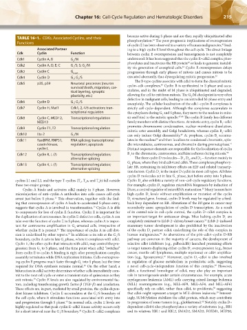
TABLE 16–1. CDKs, Associated Cyclins, and their become active during S phase and are then rapidly ubiquitinated after
41
phosphorylation. The poor prognostic implications of overexpression
Functions
of cyclin E has been observed in a variety of human malignancies, lead-
42
Associated Partner ing to a high cyclin E level throughout the cell cycle. The direct linkage
Cdk Cyclin Function between cyclin E overexpression and tumorigenesis is not completely
Cdk1 Cyclin A, B G /M understood. It has been suggested that the cyclin E–cdk2 complex phos-
2 43
Cdk2 Cyclin A, D, E; C G /S; S; G /M phorylates and inactivates the RB protein or leads to genomic instabil-
44
1 2 ity via generation of aneuploid cells. Cyclin E overexpression delays
Cdk3 Cyclin C G 0 exit progression through early phases of mitosis and causes mitosis to be
Cdk4 Cyclin D G ; G /S executed aberrantly, thus dysregulating mitotic progression. 45
1 1
Cdk5 p35, p39 Neuronal processes (neuron The B-type cyclins associate with cdk1 to form the classical mitotic
46
survival/death, migration, cor- cyclin–cdk complexes. Cyclin B is synthesized in S phase and accu-
tical layering, synaptic mulates, and in the midst of M phase is ubiquitinated and degraded,
plasticity, etc.) allowing the cell to exit from mitosis. The G /M checkpoint is very often
2
defective in malignant cells, leading to uncontrolled M-phase entry and
Cdk6 Cyclin D G ; G /S
1 1 aneuploidy. The cellular localization of the cdk1–cyclin B complexes is
Cdk7 Cyclin H, Mat1 Cdk1, 2, 4/6 activation; tran- strictly cell-cycle–dependent. Although the complexes accumulate in
scriptional regulation the cytoplasm during G and S phase, they move to the nucleus in mito-
2
Cdk8 Cyclin C, MED12, Transcriptional regulation sis and bind to the mitotic spindle. 47,48 The cyclin B family has different
MED13 family members with distinct functions. At mitotic entry, cyclin B –cdk1
1
Cdk9 Cyclin T1, T2 Transcriptional regulation promotes chromosome condensation, nuclear membrane dissolution,
mitotic aster assembly, and Golgi breakdown, whereas cyclin B –cdk1
Cdk10 Ets-2 G /M 49 2
2 can only induce Golgi disassembly. At prophase, cyclin B accumu-
1
50
Cdk11 RanBPM, RNPS1, RNA splicing; transcriptional lates in the nucleus and then localizes to condensed chromatin, spin-
casein kinase, regulation; apoptosis dle microtubules, centrosomes, and chromatin during prometaphase.
51
cyclin L Distinct sequence elements are responsible for the localization of cyclin
Cdk12 Cyclin K, L (?) Transcriptional regulation; B to the chromatin, centrosomes, and kinetochores during mitosis. 52
1
alternative splicing The three cyclin D molecules—D , D , and D —function mainly in
3
1
2
G phase, where they bind cdk4 and cdk6. These complexes phosphory-
Cdk13 Cyclin K, L (?) Transcriptional regulation; 1
alternative splicing late RB, restraining its inhibitory effects on E2F and related transcrip-
tion factors. Cyclin D is the major D cyclin in most cell types. All three
1
cyclin D molecules act in late G phase, just before entry into S phase.
1
cyclins L1 and L2, and the type T cyclins (T , T a, and T b) fall outside Cyclin D also exhibits a variety of non–cell-cycle regulatory functions.
1
2
1
2
these two major groups. For example, cyclin D regulates microRNA biogenesis by induction of
1
Cyclin A binds and activates cdk2 mainly in S phase. However, Dicer, a central regulator of microRNA maturation. Many tumors have
53
microinjection of anti–cyclin A antibodies into cells causes cell cycle high cyclin D levels without amplification or mutation of the cyclin
1
arrest just before S phase. This observation, together with the find- D structural gene. Instead, cyclin D levels may be regulated by a feed-
11
1
ing that overexpression of cyclin A leads to accelerated S-phase entry, back loop dependent on RB. Alterations of the RB gene in cancer may
suggests that cyclin A is involved in transformation. Cyclin A is able secondarily cause upregulation of cyclin D transcription. As a result
13
to compensate for loss of cyclin E function. Cyclin E is important for of its central role in cell-cycle control, the cyclin D–cdk4 complex is
the duplication of centrosomes. In cyclin E-defective cells, cyclin A can an important target for anticancer drugs. Mice lacking cyclin D are
1
54
take over the function of cyclin E in S phase, whereas cyclin A is impor- completely resistant to ErbB-2–driven breast cancer. ErbB-2–induced
tant for centrosome amplification in G -arrested cells, irrespective of mammary tumor development is also prohibited by the inactivation
2
whether cyclin E is present. The importance of cyclin A in cell divi- of the cyclin D partner cdk4, underlining the role of this complex in
36
1
55
sion is underlined by other reports. In addition to its role at the G /S human malignancies. As aberrations of the p16–cdk4–cyclin D-RB
37
1
boundary, cyclin A acts in late G phase, where it complexes with cdk1. pathway are common in the majority of cancers, the development of
2
Cyclin E, the other cyclin that interacts with cdk2, may control the pro- selective cdk4 inhibitors (e.g., palbociclib) launched promising efforts
gression from G to S phase, and the time point when cdk2 “switches” to target tumors displaying either cyclin D overexpression (e.g., breast
1
1
from cyclin E to cyclin A binding is right after prereplication complex cancer, mantle cell lymphoma, multiple myeloma) or cdk4 amplifica-
56
assembly terminates while DNA replication initiates. Cells overexpress- tion (e.g., liposarcoma). Moreover, cyclin D –cdk4 is also involved
1
ing cyclin E progress much faster through G into S phase, but the time in regulation of glucose metabolism in postmitotic cells, suggesting
1
required for DNA synthesis remains normal. On the other hand, a a novel cell-cycle–independent function of this complex. However,
57
38
bifurcation in cdk2 activity determines whether cells immediately com- cdk6, a functional homologue of cdk4, may also play an important
mit to the next cell cycle or enter a transient state of quiescence as they role in tumorigenesis under certain circumstances. For example, acute
exit mitosis. Cyclin E levels are also regulated by environmental fac- myelogenous leukemia (AML) cells carrying mixed-lineage leukemia
39
tors, including transforming growth factor-β (TGF-β) and irradiation. (MLL) rearrangements (e.g., MLL-AF9, MLL-AF4, and MLL-AF6)
58
These effects are, in part, mediated by small proteins, the cyclin-depen- specifically rely on cdk6, rather than cdk4, to proliferate, suggesting
59
dent kinase inhibitors. Cyclin E accumulates at the G /S boundary of that cdk6 might represent a target in MLL-driven leukemia. Interest-
1
the cell cycle, where it stimulates functions associated with entry into ingly, SUMOylation stabilizes the cdk6 protein, which may contribute
60
and progression through S phase. In normal cells, cyclin E levels are to progression of some tumors (e.g., glioblastoma). Notably, cyclin D–
40
highly regulated so that peak cyclin E–cdk2 kinase activity occurs only dependent cdk4/6 also phosphorylates a variety of substrates (e.g., RB1
for a short interval near the G /S boundary. Cyclin E–cdk2 complexes and its relatives RBL1 and RBL2, SMAD2, SMAD3, FOXM1, MEP50,
40
1
Kaushansky_chapter 16_p0213-0246.indd 215 9/18/15 11:56 PM