
Page 533 - Williams Hematology ( PDFDrive )
P. 533
508 Part VI: The Erythrocyte Chapter 34: Clinical Manifestations and Classification of Erythrocyte Disorders 509
TABLE 34–1. Classification of Anemia (Continued )
b. Red cell membrane disorders (Chap. 46) d. Porphyrias (congenital erythropoietic and hepatoery-
(1) Cytoskeletal membrane disorders (hereditary thropoietic porphyrias, rarely congenital erythropoi-
spherocytosis, elliptocytosis, pyropoikilocytosis) etic protoporphyria [Chap. 58])
(2) Lipid membrane disorders (hereditary abetalipo- C. Blood loss and blood redistribution
proteinemia, hereditary stomatocytosis, etc.) 1. Acute blood loss
(3) Membrane disorders associated with abnormali- 2. Splenic sequestration crisis (Chap. 56)
ties of erythrocyte antigens (McLeod syndrome, Rh
deficiency syndromes, etc.) II. Relative (increased plasma volume)
(4) Membrane disorders associated with abnormal A. Macroglobulinemia (Chap. 109)
transport (hereditary xerocytosis) B. Pregnancy (Chap. 8)
c. Red cell enzyme defects (pyruvate kinase, 5′ nucleo- C. Athletes (Chap. 33)
tidase, glucose-6-phosphate dehydrogenase deficien-
cies, other red cell enzyme disorders [Chap. 47]) D. Postflight astronauts (Chap. 33)
observed in some types of hemolytic anemia, in which the rate of red thrombopoietin, there is no evidence that the two molecules crossre-
36
cell production can be four to six times normal. In erythrocytosis, the act at the level of their respective receptors. EPO-driven erythrocytosis
37
number of red cells destroyed daily merely causes a slight increase in is generally not associated with increased platelet production.
bilirubin levels. The presence of secondary gout and splenomegaly are The increased viscosity and expansion of vascular space are
usually signs of a myeloproliferative neoplasm rather than of erythrocy- responsible for many of the signs and symptoms of polycythemia. The
tosis alone. Although considerable homology exists between EPO and characteristic rubor in patients with polycythemia vera is caused by
excessive deoxygenation of blood flowing sluggishly through dilated
cutaneous vessels. Nonspecific symptoms such as headaches, dizziness,
ERYTHROPOIESIS
tinnitus, and a reported feeling of fullness of the face and head probably
PRODUCTION DESTRUCTION are caused by a combination of increased viscosity and vascular dilata-
tion. In extreme polycythemia and some specific types of polycythemia
Stem Progenitor Precursor Mature
cell cells cells cells (e.g., methemoglobinemia; Chap. 50), cyanosis can result from greater
pool BFU-E CFU-E Erythroblasts than 4 g/dL of deoxygenated hemoglobin (accomplished more easily at
higher hemoglobin concentrations [see “blue bloaters” and “pink puff-
ers” in Chap. 57]) or greater than 1.5 g/dL of methemoglobin.
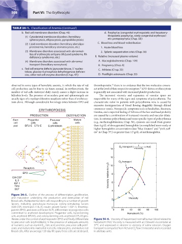
transport ( 1 x Hct )
O 2
14 viscosity
12
O transport
2
Receptors 10
EPO 8
GM-CSF Viscosity relative to H 2 O
IL-3
IGF-1 6
TPO
SCF
4
Figure 34–5. Outline of the process of differentiation, proliferation,
and maturation underlying the production and destruction of red 2 Viscosity
blood cells. Multipotential stem cells responding to a number of growth
factors, including granulocyte-monocyte colony-stimulating factors
(GM-CSF), interleukin 3 (IL-3), insulin growth factor 1 (IGF-1), thrombo- 0
poietin (TPO), and stem cell factor (SCF), differentiate to progenitor cells 0 20 40 60 80 100
committed to erythroid development. Progenitor cells, burst-forming Hematocrit (%)
unit–erythroid (BFU-E), and colony-forming unit–erythroid (CFU-E) pro-
liferate under the control of erythropoietin (EPO) and finally differentiate Figure 34–6. Viscosity of heparinized normal human blood related to
to precursor cells (erythroblasts). In the presence of adequate amounts hematocrit (Hct). Viscosity is measured with an Ostwald viscosimeter at
of nutrients, such as vitamin B , folic acid, and iron, precursor cells prolif- 37°C and expressed in relation to viscosity of saline solution. Oxygen
12
erate and mature into nucleated red cells, reticulocytes, and mature red transport is computed from Hct and O flow (1/viscosity) and is recorded
2
blood cells. After an average 120-day life span, these cells are destroyed. in arbitrary units.
Kaushansky_chapter 34_p0503-0512.indd 508 9/17/15 6:12 PM