
Page 534 - Williams Hematology ( PDFDrive )
P. 534
508 Part VI: The Erythrocyte Chapter 34: Clinical Manifestations and Classification of Erythrocyte Disorders 509
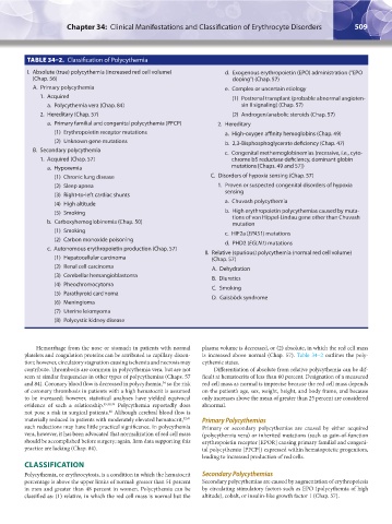
TABLE 34–2. Classification of Polycythemia
I. Absolute (true) polycythemia (increased red cell volume) d. Exogenous erythropoietin (EPO) administration (“EPO
(Chap. 56) doping”) (Chap. 57)
A. Primary polycythemia e. Complex or uncertain etiology
1. Acquired (1) Postrenal transplant (probable abnormal angioten-
a. Polycythemia vera (Chap. 84) sin II signaling) (Chap. 57)
2. Hereditary (Chap. 57) (2) Androgen/anabolic steroids (Chap. 57)
a. Primary familial and congenital polycythemia (PFCP) 2. Hereditary
(1) Erythropoietin receptor mutations a. High-oxygen affinity hemoglobins (Chap. 49)
(2) Unknown gene mutations b. 2,3-Bisphosphoglycerate deficiency (Chap. 47)
B. Secondary polycythemia c. Congenital methemoglobinemias (recessive, i.e., cyto-
1. Acquired (Chap. 57) chrome b5 reductase deficiency, dominant globin
a. Hypoxemia mutations [Chaps. 49 and 57])
(1) Chronic lung disease C. Disorders of hypoxia sensing (Chap. 57)
(2) Sleep apnea 1. Proven or suspected congenital disorders of hypoxia
(3) Right-to-left cardiac shunts sensing
(4) High altitude a. Chuvash polycythemia
(5) Smoking b. High erythropoietin polycythemias caused by muta-
tions of von Hippel-Lindau gene other than Chuvash
b. Carboxyhemoglobinemia (Chap. 50) mutation
(1) Smoking c. HIF2a (EPAS1) mutations
(2) Carbon monoxide poisoning d. PHD2 (EGLN1) mutations
c. Autonomous erythropoietin production (Chap. 57)
II. Relative (spurious) polycythemia (normal red cell volume)
(1) Hepatocellular carcinoma (Chap. 57)
(2) Renal cell carcinoma A. Dehydration
(3) Cerebellar hemangioblastoma B. Diuretics
(4) Pheochromocytoma
C. Smoking
(5) Parathyroid carcinoma D. Gaisböck syndrome
(6) Meningioma
(7) Uterine leiomyoma
(8) Polycystic kidney disease
Hemorrhage from the nose or stomach in patients with normal plasma volume is decreased, or (2) absolute, in which the red cell mass
platelets and coagulation proteins can be attributed to capillary disten- is increased above normal (Chap. 57). Table 34–2 outlines the poly-
tion; however, circulatory stagnation causing ischemia and necrosis may cythemic states.
contribute. Thrombosis are common in polycythemia vera, but are not Differentiation of absolute from relative polycythemia can be dif-
seen at similar frequencies in other types of polycythemias (Chaps. 57 ficult at hematocrits of less than 60 percent. Designation of a measured
and 84). Coronary blood flow is decreased in polycythemia, so the risk red cell mass as normal is imprecise because the red cell mass depends
34
of coronary thrombosis in patients with a high hematocrit is assumed on the patient’s age, sex, weight, height, and body frame, and because
to be increased; however, statistical analyses have yielded equivocal only increases above the mean of greater than 25 percent are considered
evidence of such a relationship. 35,38,39 Polycythemia reportedly does abnormal.
not pose a risk in surgical patients. Although cerebral blood flow is
40
materially reduced in patients with moderately elevated hematocrit, 32,41 Primary Polycythemias
such reductions may have little practical significance. In polycythemia Primary or secondary polycythemias are caused by either acquired
vera, however, it has been advocated that normalization of red cell mass (polycythemia vera) or inherited mutations (such as gain-of-function
should be accomplished before surgery; again, firm data supporting this erythropoietin receptor [EPOR] causing primary familial and congeni-
practice are lacking (Chap. 84). tal polycythemia [PFCP]) expressed within hematopoietic progenitors,
leading to increased production of red cells.
CLASSIFICATION
Polycythemia, or erythrocytosis, is a condition in which the hematocrit Secondary Polycythemias
percentage is above the upper limits of normal: greater than 51 percent Secondary polycythemias are caused by augmentation of erythropoiesis
in men and greater than 48 percent in women. Polycythemia can be by circulating stimulatory factors such as EPO (polycythemia of high
classified as: (1) relative, in which the red cell mass is normal but the altitude), cobalt, or insulin-like growth factor 1 (Chap. 57).
Kaushansky_chapter 34_p0503-0512.indd 509 9/17/15 6:12 PM