
Page 530 - Williams Hematology ( PDFDrive )
P. 530
504 Part VI: The Erythrocyte Chapter 34: Clinical Manifestations and Classification of Erythrocyte Disorders 505
120 Murmurs usually are heard during systole. Murmurs and bruits
have been described in many regions, such as over the jugular vein,
110 the closed eye, and the parietal region of the skull, and may be sensed by
the patient as roaring in the ears (tinnitus), especially at night. They disap-
Total blood volume (mL/kg) 90 The myocardium tolerates a prolonged period of sustained hyperactiv-
pear promptly after the hemoglobin concentration is restored to normal.
100
20
ity. However, angina pectoris and high-output failure may supervene if
anemia is so severe that it exceeds myocardial oxygen demands or if the
80
patient has coronary artery disease. Cardiomegaly, pulmonary conges-
tion, ascites, and edema have been observed, and they require prompt
70
treatment with oxygen and transfusion of packed red cells.
60
Increased Pulmonary Function
50 Significant anemia leads to a compensatory increase in respiratory rate
that decreases the oxygen gradient from ambient air to alveolar air and
40 increases the amount of oxygen available to oxygenate a greater than
0 20 40 60 80
normal cardiac output. Consequently, exertional dyspnea and orthopnea
Hematocrit (%)
are characteristic clinical manifestations of moderate to severe anemia. 19–22
Figure 34–3. Relationship between hematocrit and total blood volume
in normal individuals and in patients with anemia and polycythemia. Increased Red Cell Production
(Reproduced with permission from Huber H, Lewis SM and Szur L. The The most appropriate response to anemia is a compensatory increase
Influence of Anaemia, Polycythaemia and Splenomegaly on the Relationship of red cell production, which may increase about twofold to threefold
between Venous Haematocrit and Red-Cell Volume. Br J Haematol acutely and fourfold to sixfold chronically, and 10-fold in the most
10:567–575,1964.) extreme case. The increase is mediated by increased production of EPO.
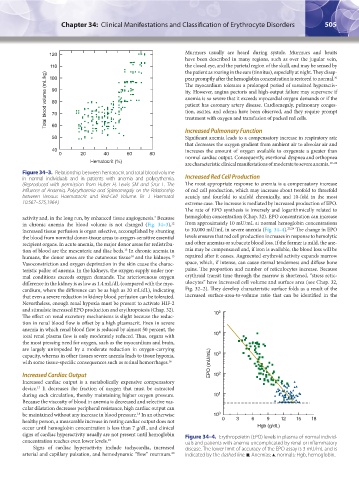
The rate of EPO synthesis is inversely and logarithmically related to
activity and, in the long run, by enhanced tissue angiogenesis. Because hemoglobin concentration (Chap. 32). EPO concentration can increase
2
in chronic anemia the blood volume is not changed (Fig. 34–3), from approximately 10 mU/mL at normal hemoglobin concentrations
12
increased tissue perfusion is organ selective, accomplished by shunting to 10,000 mU/mL in severe anemia (Fig. 34–4). 23,24 The change in EPO
the blood from nonvital donor-tissue areas to oxygen-sensitive essential levels ensures that red cell production increases in response to hemolytic
recipient organs. In acute anemia, the major donor areas for redistribu- and other anemias or subacute blood loss. If the former is mild, the ane-
tion of blood are the mesenteric and iliac beds. In chronic anemia in mia may be compensated and, if iron is available, the blood loss will be
13
humans, the donor areas are the cutaneous tissue and the kidneys. repaired after it ceases. Augmented erythroid activity expands marrow
14
15
Vasoconstriction and oxygen deprivation in the skin cause the charac- space, which, if intense, can cause sternal tenderness and diffuse bone
teristic pallor of anemia. In the kidneys, the oxygen supply under nor- pains. The proportion and number of reticulocytes increase. Because
mal conditions exceeds oxygen demands. The arteriovenous oxygen erythroid transit time through the marrow is shortened, “stress retic-
difference in the kidney is as low as 1.4 mL/dL (compared with the myo- ulocytes” have increased cell volume and surface area (see Chap. 32,
cardium, where the difference can be as high as 20 mL/dL), indicating Fig. 32–2). They develop characteristic surface folds as a result of the
that even a severe reduction in kidney blood perfusion can be tolerated. increased surface-area-to-volume ratio that can be identified in the
Nevertheless, enough renal hypoxia must be present to activate HIF-2
and stimulate increased EPO production and erythropoiesis (Chap. 32). 5
The effect on renal excretory mechanisms is slight because the reduc- 10
tion in renal blood flow is offset by a high plasmacrit. Even in severe
anemia in which renal blood flow is reduced by almost 50 percent, the
total renal plasma flow is only moderately reduced. Thus, organs with 10 4
the most pressing need for oxygen, such as the myocardium and brain,
are largely unimpeded by a moderate reduction in oxygen-carrying
capacity, whereas in other tissues severe anemia leads to tissue hypoxia, 10 3
with some tissue-specific consequences such as retinal hemorrhages. 16 EPO (mU/mL)
Increased Cardiac Output 10 2
Increased cardiac output is a metabolically expensive compensatory
device. It decreases the fraction of oxygen that must be extracted
17
during each circulation, thereby maintaining higher oxygen pressure. 10 1
Because the viscosity of blood in anemia is decreased and selective vas-
cular dilatation decreases peripheral resistance, high cardiac output can
be maintained without any increase in blood pressure. In an otherwise 10 0
18
healthy person, a measurable increase in resting cardiac output does not 0 0 3 6 9 12 15 18
occur until hemoglobin concentration is less than 7 g/dL, and clinical Hgb (g/dL)
signs of cardiac hyperactivity usually are not present until hemoglobin Figure 34–4. Erythropoietin (EPO) levels in plasma of normal individ-
concentration reaches even lower levels. 19 uals and patients with anemia uncomplicated by renal or inflammatory
Signs of cardiac hyperactivity include tachycardia, increased disease. The lower limit of accuracy of the EPO assay is 3 mU/mL and is
arterial and capillary pulsation, and hemodynamic “flow” murmurs. indicated by the dashed line. •, Anemias; ▲, normals; Hgb, hemoglobin.
20
Kaushansky_chapter 34_p0503-0512.indd 505 9/17/15 6:12 PM