
Page 555 - Williams Hematology ( PDFDrive )
P. 555
530 Part VI: The Erythrocyte Chapter 35: Aplastic Anemia: Acquired and Inherited 531
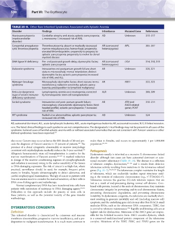
TABLE 35–9. Other Rare Inherited Syndromes Associated with Aplastic Anemia
Disorder Findings Inheritance Mutated Gene References
Ataxia-pancytopenia Cerebellar atrophy and ataxia; aplastic pancytopenia; AD Unknown 315–317
(myelocerebellar ± monosomy 7; increased risk of AML
disorder)
Congenital amegakaryo- Thrombocytopenia; absent or markedly decreased AR (compound MPL 305–307
cytic thrombocytopenia marrow megakaryocytes; hemorrhagic propensity; heterozygotes)
elevated thrombopoietin; propensity to progress to
aplastic pancytopenia; propensity to evolve to clonal
myeloid disease
DNA ligase IV deficiency Pre- and postnatal growth delay; dysmorphic facies; AR (compound LIG4 314, 318, 319
aplastic pancytopenia heterozygotes)
Dubowitz syndrome Intrauterine and postpartum growth failure; short AR Unknown 320, 321
stature; microcephaly; mental retardation; distinct
dysmorphic facies; aplastic pancytopenia; increased
risk of AML and ALL
Nijmegen breakage Microcephaly; dystrophic facies; short stature; immu- AR NBS1 322, 323
syndrome nodeficiency; radiation sensitivity; aplastic pancy-
topenia; predisposition to lymphoid malignancy
Reticular dysgenesis Lymphopenia; anemia and neutropenia; corrected XLR Unknown 308, 309
(type of severe immuno- by hematopoietic stem cell transplantation
deficiency syndrome)
Seckel syndrome Intrauterine and post- partum growth failure; AR ATR (and 310–314
microcephaly; characteristic dysmorphic facies (bird- RAD3-related
headed profile); aplastic pancytopenia; ? increased gene); PCNT
risk of AML
WT syndrome Radial/ulnar abnormalities; aplastic pancytopenia; AD Unknown 324
increased risk of AML
AD, autosomal dominant; ALL, acute lymphocytic leukemia; AML, acute myelogenous leukemia; AR, autosomal recessive; XLR, X-linked recessive.
note: The listed clinical findings in each syndrome are not comprehensive. The designated clinical findings may not be present in all cases of the
syndrome. Isolated cases of familial aplastic anemia with or without associated anomalies that are not consistent with Fanconi anemia or other
defined syndromes have been reported. 227
also occur. Cancers may occur as late as the fifth decade of life and pre- males than in females, and occurs in approximately 1 per 1,000,000
265
cede the diagnosis of Fanconi anemia in 25 percent of patients. The population. 251,272
presence of a clonal cytogenetic abnormality or marrow morphology
consistent with myelodysplasia markedly reduces the 5-year survival. Pathogenesis
232
Allogeneic hematopoietic stem cell transplantation is curative for the Dyskeratosis usually is inherited as a recessive X-chromosome–linked
marrow manifestations of Fanconi anemia. 266–269 A marked reduction disorder although rare cases can have autosomal dominant or auto-
in dosage of the marrow-conditioning regimen of cyclophosphamide somal recessive inheritance (Table 35–10). The disease is a reflection
and radiation is necessary owing to the undue sensitivity of the tissues of telomere complex dysfunction, 273–276 and it results from defective
to DNA-damaging exposures. The risk of cancer is so high that, where telomerase activity resulting from mutations in the telomerase-related
practical, surveillance should be used; for example, frequent pelvic genes (Fig. 35–7). 274,278,279 The telomerase complex maintains the length
exams in females, hepatic ultrasonography to detect adenomas, and of telomeres, which are nucleotide tandem repeat structures resid-
careful oropharyngeal examinations. Therapy of cancer in patients with ing at the termini of eukaryotic chromosomes (e.g., 5′-TTAGGG-3′).
Fanconi anemia needs to consider the marked sensitivity of their cells to Telomerase restores the guanine (G)-rich telomere repeats that are
DNA cross-linking agents and radiotherapy. lost as a result of end-processing during normal cell division. Com-
Normal complementary DNA has been transferred into cells from bined with protein, located at the ends of chromosomes, they maintain
patients with restoration of resistance to DNA damaging agents. 270,271 chromosome integrity by preventing end-to-end chromosome fusion,
Difficulties in this approach include the paucity of stem cells in preventing chromosome degradation, and preventing chromosome
these patients, as well as the potential toxicity of the gene transfer instability. In dyskeratosis congenita, the telomeres are markedly short-
methodology. ened resulting in genomic instability and cell (including marrow cell)
apoptosis, and the underlying gene defects may alter Box H/ACA small
DYSKERATOSIS CONGENITA nucleolar RNAs, such as the telomerase RNA component, TERC, that
is central to telomere maintenance.
Rapidly proliferating cells are at
279a
Definition highest risk for dysfunction. Mutations of the DKC1 gene are respon-
This inherited disorder is characterized by cutaneous and mucous sible for the X-linked recessive form. DKC1 encodes dyskerin, which
membrane abnormalities, progressive marrow insufficiency, and a pre- is a conserved multifunctional protein component of the telomerase
disposition to malignant transformation. It is much more common in complex. Mutations of the TERT, TERC, and TINF2 genes are the
Kaushansky_chapter 35_p0513-0538.indd 530 9/19/15 12:24 AM