
Page 57 - Williams Hematology ( PDFDrive )
P. 57
32 Part I: Clinical Evaluation of the Patient Chapter 3: Examination of the Marrow 33
Storage Diseases only on biopsy specimens, necessitates examination by special stains
Storage disorders, such as Gaucher and Niemann-Pick diseases (Chap. for fungal and mycobacterial organisms, but the differential diagnosis
72), are characterized by abnormal macrophages containing stored is extensive. 53,61
material in various forms seen in aspirate or biopsy. Reactive cells,
53
such as the histiocytes with “sea-blue” inclusion granules (Chap. 72) or
pseudo-Gaucher cells associated with chronic myelogenous leukemia NECROSIS AND GELATINOUS
(CML) (Chap. 89), can resemble the cells seen in storage disorders. TRANSFORMATION
53
Marrow necrosis may occur in a variety of disorders, particularly sickle
Amyloidosis cell disease and neoplastic processes involving the marrow. Aspirates
62
Amyloid refers to extracellular proteins that become insoluble as a result of necrotic marrow stained with polychrome stains contain cells with
of alteration in secondary structure to form beta pleated sheets. Amy- indistinct margins and smudged basophilic nuclei surrounded by aci-
loid light-chain amyloidosis may result from a plasma cell neoplasm, dophilic material. Marrow sections stained with hematoxylin and eosin
and is associated with nephrotic syndrome, restrictive cardiomyopa- show loss of normal marrow architecture, indistinct cellular margins,
thy, neuropathy and other tissue involvement. Amyloid deposits can be and a background of amorphous eosinophilic material. Patients with
identified in the marrow by characteristic birefringence or fluorescence severe weight loss may develop gelatinous transformation of the mar-
of deposits when stained with Congo Red. 54 row, characterized by amorphous extracellular material (proteogly-
cans), fat atrophy, and marrow hypoplasia. 63
INFECTIONS
Infectious organisms with an intracellular location, such as Leishma- MORPHOLOGIC DIFFERENTIATION OF
nia, Histoplasma, and Toxoplasma, can be visualized in monocytic
55
cells by morphologic examination of the marrow (Fig. 3–3). Identifi- HEMATOPOIETIC LINEAGES
cation of mycobacterial organisms in the marrow by acid-fast staining
lacks sensitivity but allows early diagnosis in one-third of cases with OVERVIEW
HIV-related Mycobacterium avium complex infection. Microscopic Marrow aspirate films should be examined under low-power magnifi-
56
examination and culture of the marrow are the most sensitive diagnos- cation to assess the cellularity of particles and estimate the number of
tic tests for disseminated leishmaniasis. Mycobacteria, also, may be megakaryocytes, plasma cells, and mast cells. Low-power examination
57
cultured from marrow. Marrow morphology also is a sensitive diagnos- may also permit detection of malignancy or abnormal storage cells.
tic tool for detecting disseminated histoplasmosis. However, marrow The entire film should be examined, including the particles, and higher
58
culture has a low diagnostic yield in the workup of fever of unknown magnification should be used to study any abnormalities discovered.
origin in nonimmunosuppressed patients. Definitive diagnoses aris- Similarly, biopsy sections are examined at low power to assess adequacy,
59
ing from marrow examination in this setting are usually hematologic overall cellularity, presence of infiltrative disease, and cellularity of the
malignancies. 59,60 The presence of marrow granulomas, recognizable major hematopoietic lineages.
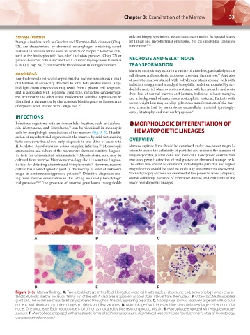
A B C
D E F
Figure 3–3. Marrow findings. A. Two osteoblasts are in this field. Elongated ovoid cells with nucleus at extreme end, a morphology which charac-
teristically looks like the nucleus is falling out of the cell. A clear area is apparent spaced at an interval from the nucleus. B. Osteoclast. Multinucleated
giant cell. The nuclei are characteristically scattered throughout the cell, appearing separate. C. Macrophage (arrow), relatively large cell with circular
nucleus and abundant cytoplasm. Ingested debris and few vacuoles. D. Macrophage (two). Prussian blue stain. Relatively large cell with circular
nuclei. One binucleate. Each macrophage is full of iron as indicated by blue reaction product of stain. E. Macrophage engorged with Histoplasma cap-
sulatum. F. Macrophage engorged with amastigote forms of Leishmania donovani. (Reproduced with permission from Lichtman’s Atlas of Hematology,
www.accessmedicine.com.)
Kaushansky_chapter 03_p0027-0040.indd 33 17/09/15 5:38 pm