
Page 689 - Williams Hematology ( PDFDrive )
P. 689
664 Part VI: The Erythrocyte Chapter 46: Erythrocyte Membrane Disorders 665
The membrane proteins are classified as either integral or periph-
eral based on the ease with which they can be removed from whole red
cell membrane preparations in the laboratory. Integral or transmem-
brane proteins are embedded in the lipid bilayer by hydrophobic inter-
actions and require detergents to extract them. They often protrude 642
from the bilayer and extend into the plasma and/or the interior of the Asn
erythrocyte and these structural features correlate with their functions
as transport proteins, receptors, signaling molecules, and carriers of red
cell antigens.
Peripheral proteins constitute the membrane skeleton and are 1 2 3 4 5 67 89 10 11 12 13
loosely attached to the cytoplasmic face of the lipid bilayer and can be
extracted by high or low salt concentrations or by high pH. Attachment 400
is mediated indirectly by covalent or noncovalent interactions with the Carbonic
cytoplasmic domains of the transmembrane proteins, as well as by direct anhydrase
interactions with the inner leaflet of the lipid bilayer. These associations C 911
are dynamic and the affinity of binding is regulated by post-translational
modifications of the proteins, including phosphorylation, methylation,
glycosylation, or lipid modification (myristoylation, palmitoylation, or Cytoplasmic protein binding domain:
farnesylation). Peripheral proteins typically function either as structural Ankyrin, adducin, 4.1R, 4.2
proteins and form part of the membrane skeleton or they serve as linker Glycolytic enzymes
proteins attaching the skeleton to the bilayer. Hemoglobin, hemichromes
Many erythrocyte proteins belong to superfamilies and have
homologues in nonerythroid cells that are structurally related but are N
encoded by different genes. This genetic diversity explains why the clini- 1 8
cal expression of most (but not all) red cell membrane protein mutations Tyr-P
is confined to the erythroid lineage. Several proteins exist in different
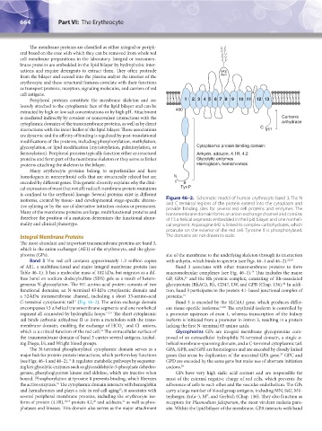
isoforms, created by tissue- and developmental stage-specific alterna- Figure 46–2. Schematic model of human erythrocyte band 3. The N
tive splicing or by the use of alternative initiation codons or promoters. and C terminal regions of the protein extend into the cytoplasm and
provide binding sites for several red cell proteins and enzymes. The
Many of the membrane proteins are large, multifunctional proteins and transmembrane domain forms an anion exchange channel and consists
therefore the position of a mutation determines the functional abnor- of 13 α helical segments embedded in the lipid bilayer and one nonheli-
mality and clinical phenotype. cal segment. Asparagine 642 is linked to complex carbohydrates, which
protrude on the exterior of the red cell. Tyrosine 8 is phosphorylated.
Integral Membrane Proteins The domains are not drawn to scale.
The most abundant and important transmembrane proteins are band 3,
which is the anion exchanger (AE1) of the erythrocyte, and the glyco-
phorins (GPs). site of the membrane to the underlying skeleton through its interaction
Band 3 The red cell contains approximately 1.2 million copies with ankyrin, which binds to spectrin (see Figs. 46–1 and 46–2). 22,23
of AE1, a multifunctional and major integral membrane protein (see Band 3 associates with other transmembrane proteins to form
Table 46–1). It has a molecular mass of 102 kDa, but migrates as a dif- macromolecular complexes (see Fig. 46–1). This includes the major
24
fuse band on sodium dodecylsulfate (SDS) gels as a result of hetero- GP, GPA, and the Rh protein complex, consisting of Rh-associated
25
geneous N-glycosylation. The 911-amino-acid protein consists of two glycoprotein (RhAG), Rh, CD47, LW, and GPB (Chap. 136). In addi-
24
functional domains; an N-terminal 43-kDa cytoplasmic domain and tion, band 3 participates in the protein 4.1-based junctional complex of
a 52-kDa transmembrane channel, including a short 33-amino-acid proteins. 19
C-terminal cytoplasmic tail (Fig. 46–2). The anion exchange domain Band 3 is encoded by the SLC4A1 gene, which produces differ-
11
encompasses 13 α helical transmembrane segments and one nonhelical ent tissue-specific isoforms. 11,26 The erythroid isoform is controlled by
segment all connected by hydrophilic loops. 12,13 The short cytoplasmic a promoter upstream of exon 1, whereas transcription of the kidney
tail binds carbonic anhydrase II to form a metabolon with the trans- isoform is initiated from a promoter in intron 3, resulting in a protein
membrane domain, enabling the exchange of HCO and Cl anions, lacking the first N-terminal 65 amino acids.
–
–
3
which is a critical function of the red cell. The extracellular surface of Glycophorins GPs are integral membrane glycoproteins com-
14
the transmembrane domain of band 3 carries several antigens, includ- posed of an extracellular hydrophilic N-terminal domain, a single α-
ing Diego, I/i, and Wright blood groups. helical membrane-spanning domain, and a C-terminal cytoplasmic tail.
The N-terminal phosphorylated cytoplasmic domain serves as a GPA, GPB, and GPE are homologous and are encoded by closely linked
major hub for protein-protein interactions, which perform key functions genes that arose by duplication of the ancestral GPA gene. GPC and
27
(see Figs. 46–1 and 46–2). It regulates metabolic pathways by sequester- GPD are encoded by the same gene but make use of alternate initiation
15
ing key glycolytic enzymes such as glyceraldehyde-3-phosphate dehydro- codons. 28
genase, phosphoglycerate kinase and aldolase, which are inactive when GPs have very high sialic acid content and are responsible for
bound. Phosphorylation at tyrosine 8 prevents binding, which liberates most of the external negative charge of red cells, which prevents the
the active enzymes. The cytoplasmic domain interacts with hemoglobin adherence of cells to each other and the vascular endothelium. The GPs
16
and hemichromes and plays a role in red cell aging ; it associates with carry a large number of blood group antigens, including MN, SsU, Mil-
17
several peripheral membrane proteins, including the erythrocyte iso- tenberger, En(a–), M , and Gerbich (Chap. 136). They also function as
K
20
21
form of protein (4.1R), 18,19 protein 4.2, and adducin, as well as phos- receptors for Plasmodium falciparum, the most virulent malaria para-
phatases and kinases. This domain also serves as the major attachment site. Within the lipid bilayer of the membrane, GPA interacts with band
Kaushansky_chapter 46_p0661-0688.indd 664 9/17/15 6:41 PM