
Page 722 - Williams Hematology ( PDFDrive )
P. 722
696 Part VI: The Erythrocyte Chapter 47: Erythrocyte Enzyme Disorders 697
+
140
+
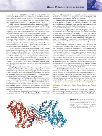
requires the presence of NADP (Fig. 47–5). Hence, NADP is bound Deficiency of the enzyme has been described as one of the rarest human
169
to the enzyme both as a structural component, in the subunit interface, disorders. It manifests with progressive leukoencephalopathy and
and as one of the substrates of the reaction. 141–143 Under physiologic con- neuropathy. No dysfunction of red cells was reported. 170
ditions, the active human enzyme exists in a dimer–tetramer equilib- Ribulose-Phosphate Epimerase Ribulose-phosphate epimerase
rium. Lowering the pH causes a shift toward the tetrameric form. 141,144,145 converts ribulose-5-phosphate to xylulose-5-phosphate. The exact
146
G6PD is strongly inhibited by physiologic amounts of NADPH activity of this enzyme in human hemolysates has not been reported but
147
and, to a lesser extent, by physiologic concentrations of ATP. It has seems to be less than that of ribosephosphate isomerase.
much higher enzyme-activity in reticulocytes than in mature red cells, Transketolase Transketolase effects the transfer of two carbon
especially for the mutant forms of the enzyme. 43,148 This may complicate atoms from xylulose-5-phosphate to ribose-5-phosphate, resulting in
diagnosing G6PD deficiency in patients with high reticulocyte counts the formation of the 7-carbon sugar sedoheptulose-7-phosphate and the
following a hemolytic episode (see “Laboratory Features” below). 3-carbon sugar glyceraldehyde-3-phosphate. 171–173 It can also catalyze
The three-dimensional model of the crystal structure of human the reaction between xylulose-5-phosphate and erythrose-4-phosphate,
G6PD shows that the G6PD monomer is built up by two domains, a producing fructose-6-phosphate and glyceraldehyde-3-phosphate. Thi-
N-terminal domain and a large β + α domain with an antiparallel nine- amine pyrophosphate is a coenzyme for transketolase, and the activity
stranded sheet. The extensive interface between the two monomers is of erythrocyte transketolase is used as an index of the adequacy of thi-
of crucial importance for enzymatic stability and activity. The fully amine nutrition. 174
143
conserved amino acids 198 to 205 (Arg-Ile-Asp-His-Tyr-Leu-Gly-Lys) Transaldolase The conversion of seduhepulose-7-phosphate and
are essential for substrate binding and catalysis. 143,149–151 glyceraldehyde-3-phosphate into erythrose-4-phosphate and fruc-
175
The G6PD gene is located on the X-chromosome (Xq28). It spans tose-6-phosphate is catalyzed by transaldolase. This is another one in
18 kb, and consists of 13 exons of which exon 1 is noncoding. Methy- the series of molecular rearrangements that leads in the conversion of the
lation of certain cytidines at the 3′ end is believed to have a regulatory 5-carbon sugar formed in the phosphogluconate dehydrogenase step to
function. 152,153 The 3′-UTR (untranslated region) also harbors putative metabolic intermediates of the EMP. Transaldolase deficiency was first
microRNA (miRNA) target sites, which potentially could have a func- reported in 2001 as a new inborn error of the pentose phosphate path-
176
tional effect on the downregulation of G6PD mRNA, thereby affecting way. To date, 23 patients from 13 families have been described. 177,178
the stability of G6PD mRNA and translation, or the miRNA regulation It is a pleiotropic metabolic disorder, and patients present in the neona-
process. G6PD deficiency is one of the world’s most common hered- tal or antenatal period with dysmorphic features, hepatosplenomegaly,
154
itary disorders. Many mutations and variants have been reported and abnormal liver function, cardiac defects, thrombocytopenia, bleeding
studied. 1,2,155–160 tendencies, and anemia. The latter appears to be hemolytic in nature,
Phosphogluconolactonase Although 6-phosphogluconolactone possibly because of decreased levels of NADPH. 179,180
is the direct product of the oxidation of glucose-6-phosphate by G6PD l-Hexonate Dehydrogenase Red cells contain L-hexonate dehy-
and hydrolyzes spontaneously at a physiologic pH, enzymatic hydrolysis drogenase, an enzyme that has the capacity to reduce aldoses such as
is much more rapid and is required for normal metabolic flow through glucose, galactose, or glyceraldehyde to their corresponding polyol (i.e.,
the stimulated hexose monophosphate pathway. 161,162 Partial deficiency glucose to sorbitol, galactose to dulcitol, and glyceraldehyde to glyc-
181
163
of the enzyme has been observed and is probably benign. 164 erol). NADPH serves as a hydrogen donor for this reaction. Aldose
Phosphogluconate Dehydrogenase Phosphogluconate dehy- reductase is another enzyme that can catalyze this reaction. It is pres-
182
drogenase catalyzes the oxidation of phosphogluconate to ribulose-5- ent in red cells, and increased levels have been implicated in diabetic
+
phosphate and CO and the reduction of NADP to NADPH. Variability complications, such as retinopathy and autonomic neuropathy. 184
183
2
of electrophoretic mobility of the enzyme is common in humans and in
several animal species. Deficiency of the enzyme has been observed Utilization of Substrates Other Than Glucose as Energy
165
only rarely and appears to be essentially innocuous or possibly associ- Sources
ated with mild hemolysis. 166–168 The red cell has the capacity to use several other substrates in addition to
Ribosephosphate Isomerase Ribosephosphate isomerase catalyzes glucose as a source of energy. Among these are adenosine, inosine, fruc-
the interconversion of ribulose-5-phosphate and ribose-5-phosphate. tose, mannose, galactose, dihydroxyacetone, and lactate. Although in
185
Figure 47–5. A dimer of human glucose-6-
phosphate dehydrogenase. Subunits A and
B are colored red and blue. Structural nico-
tinamide adenine dinucleotide phosphate
+
(NADP ) molecules are drawn in ball-and-
stick mode and colored dark blue.
Kaushansky_chapter 47_p0689-0724.indd 697 9/17/15 6:44 PM