
Page 721 - Williams Hematology ( PDFDrive )
P. 721
696 Part VI: The Erythrocyte Chapter 47: Erythrocyte Enzyme Disorders 697
The reaction is facilitated by the presence of metal ions. Aside from Both intra- and intersubunit interactions are considered to be key deter-
113
its enzymatic function in the glycolytic pathway, α-enolase (ENO1) has minants of the allosteric response, which involves switching of the PK
been implicated in numerous diseases, including metastatic cancer, tetramer from the low-affinity T state to the high-affinity R state. 128–132
114
autoimmune disorders, ischemia, and bacterial infection. The gene Red cell PK manifests sigmoid kinetics with respect to phosphoenolpy-
for enolase (ENO1) is located on chromosome 1p36.23. Enolase defi- ruvate in the absence of fructose-1,6-diphosphate. Hyperbolic kinetics
ciency is extremely rare. Although it has been reported in association are observed in the presence of even minute amounts of fructose-1,6-
with hereditary nonspherocytic hemolytic anemia, 115,116 a clear cause- diphosphate, 122,133 so that at low concentrations of phosphoenolpyru-
and-effect relationship has not yet been firmly established. vate the enzyme activity is greatly increased by fructose diphosphate.
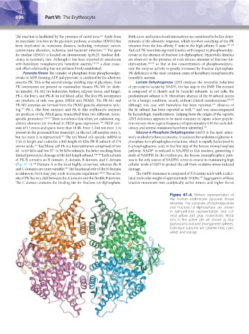
Pyruvate Kinase The transfer of phosphate from phosphoenolpy- PK deficiency is the most common cause of hereditary nonspherocytic
ruvate to ADP, forming ATP and pyruvate, is catalyzed by the allosteric hemolytic anemia.
enzyme PK. This is the second energy-yielding step of glycolysis. Four Lactate Dehydrogenase LDH catalyzes the reversible reduction
PK isoenzymes are present in mammalian tissues: PK-M1 (in skele- of pyruvate to lactate by NADH, the last step in the EMP. The enzyme
tal muscle), PK-M2 (in leukocytes, kidney, adipose tissue, and lungs), is composed of H (heart) and M (muscle) subunits. In red cells, the
PK-L (in liver), and PK-R (in red blood cells). The four PK isoenzymes predominant subunit is H. Hereditary absence of the H subunit seems
are products of only two genes (PKLR and PKM2). The PK-M1 and to be a benign condition, usually without clinical manifestations, 134,135
136
PK-M2 enzymes are formed from the PKM2 gene by alternative splic- although one case with hemolysis has been reported. Absence of
137
ing. PK-L (the liver enzyme) and PK-R (the erythrocyte enzyme) the M subunit has been reported as well, and was unaccompanied
117
are products of the PKLR gene, transcribed from two different, tissue- by hematologic manifestations. Judging from the origin of the reports,
specific promoters. 118,119 There is evidence that other, yet unknown, reg- LDH deficiency appears to be most common in Japan, where popula-
ulatory elements are involved in PKLR gene expression. PKLR con- tion surveys show a gene frequency of approximately 0.05 for each defi-
120
sists of 12 exons and spans more than 10 kb. Exon 2, but not exon 1, is ciency, and several mutations have been identified. 138
present in the processed liver transcript; in the red cell enzyme exon 1, Glucose-6-Phosphate Dehydrogenase G6PD is the most exten-
but not exon 2, is represented. The red blood cell–specific mRNA is sively studied erythrocyte enzyme. It catalyzes the oxidation of glucose-6-
119
2 kb in length and codes for a full-length 63-kDa PK-R subunit of 574 phosphate to 6-phosphogluconolactone, which is rapidly hydrolyzed to
121
amino acids. Red blood cell PK is a heterotetramer comprised of two 6-phosphogluconic acid, in the first step of the hexose monophosphate
62- to 63-kDa and two 57- to 58-kDa subunits, the latter resulting from pathway. NADP is reduced to NADPH in this reaction, generating 1
+
limited proteolytic cleavage of the full-length subunit. 122,123 Each subunit mole of NADPH. In the erythrocyte, the hexose monophosphate path-
of PK-R contains an N domain, A domain, B domain, and C domain way is the only source of NADPH, which is crucial in maintaining high
(Fig. 47–4). Domain A is the most highly conserved, whereas the B cellular levels of GSH to protect the cell from oxidative stress-induced
124
and C domains are more variable. The functional role of the N domain damage.
125
is unknown, but it may play a role in enzyme regulation. 126,127 The active The G6PD monomer is composed of 515 amino acids with a calcu-
139
site of PK lies in a cleft between the A domain and the flexible B domain. lated molecular weight of approximately 59 kDa. Aggregation of these
The C domain contains the binding site for fructose-1,6-diphosphate. inactive monomers into catalytically active dimers and higher forms
Figure 47–4. Ribbon representation of
the human erythrocyte pyruvate kinase
tetramer. The substrate phosphoglycolate
and fructose-1,6-diphosphate are shown
in ball-and-stick representation, and col-
ored yellow and gray, respectively. Metal
ions in the active site are shown as blue
(potassium) and pink (manganese) spheres.
Individual subunits are colored lime, cyan,
violet, and orange.
Kaushansky_chapter 47_p0689-0724.indd 696 9/17/15 6:44 PM