
Page 719 - Williams Hematology ( PDFDrive )
P. 719
694 Part VI: The Erythrocyte Chapter 47: Erythrocyte Enzyme Disorders 695
reaction is exquisitely sensitive to fructose 1,6-diphosphate, the prod- that has been made available by the oxidation of NADPH. NADPH
uct of PFK. Conversely, other metabolic products may serve as strong appears to function primarily as a substrate for the reduction of
enzyme inhibitors. In addition, there is increasing evidence that glyco- glutathione-containing disulfides in the erythrocyte through mediation
lytic enzymes assemble into enzyme complexes to the interior of the red of the enzyme glutathione reductase, which catalyzes the conversion
cell membrane. The assembly of these complexes seems to be regulated of oxidized glutathione (GSSG) to reduced glutathione (GSH) and the
37
by the oxygen status of hemoglobin and the phosphorylation status of reduction of mixed disulfides of hemoglobin and GSH. 42
band 3, 37,38 suggesting they play a direct role in the regulation of oxygen- Enzymes of Glucose Metabolism
dependent changes in glycolytic and pentose shunt fluxes. 39 Hexokinase Hexokinase catalyzes the phosphorylation of glucose
Notably, a number of glycolytic enzymes show additional func- in position 6 by ATP. It thus serves as the first step in the utilization of
tional activities. For instance, in addition to its role in glycolysis, glu- glucose, whether by the anaerobic or the hexose monophosphate path-
cosephosphate isomerase also functions as a neuroleukin or autocrine way. Mannose or fructose may also serve as a substrate for this enzyme.
motility factor. Another example is enolase, that has been reported to Hexokinase is the glycolytic enzyme with the lowest activity. Reticulo-
also function as plasminogen receptor. 40,41 The additional functional cytes have much higher levels of hexokinase activity than do mature red
activities of these “moonlighting” enzymes could contribute to the com- cells. 43,44
plexity of the phenotype of the associated disorder. Hexokinase has an absolute requirement for magnesium. It is
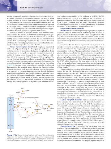
Hexose Monophosphate Shunt Not all the glucose metabolized strongly inhibited by its product, glucose-6-phosphate, and is released
by the red cell passes through the direct glycolytic pathway. A direct from this inhibition by the inorganic phosphate ion 45,46 and by high
47
oxidative pathway of metabolism, the hexose monophosphate shunt, concentrations of glucose. Inorganic phosphate enhances the rate of
also functions. In this pathway, glucose-6-phosphate is oxidized at glucose utilization by red cells. This effect is not exerted through hexoki-
position 1, yielding carbon dioxide. In the process of glucose oxida- nase but through stimulation of the PFK reaction, resulting in a lowered
+
tion, NADP is reduced to NADPH (reduced NAD phosphate). The glucose-6-phosphate concentration within the cell and thus releasing
49
48
pentose phosphate formed when glucose is decarboxylated undergoes hexokinase from inhibition. GSSG and other disulfides, as well as
50
a series of molecular rearrangements, eventuating in the formation of a 2,3-BPG, inhibit hexokinase. The determination of the structures
triose, glyceraldehyde-3-phosphate, and a hexose, fructose-6-phosphate of the human and rat hexokinase isozymes have provided substantial
(Fig. 47–3). These are normal intermediates in anaerobic glycolysis and insight into ligand-binding sites and subsequent modes of interaction
thus can rejoin that metabolic stream. Because the glucose phosphate of these ligands. 51,52
isomerase reaction is freely reversible, allowing fructose-6-phosphate The two major fractions of red cell hexokinase (HK) have been des-
to be converted to glucose-6-phosphate, recycling through the hexose ignated HK and HK ; the latter fraction being unique to erythrocytes
R
I
monophosphate pathway is also possible. Unlike the anaerobic glyco- and particularly to reticulocytes. Both red cell isozymes are monomers
53
lytic pathway, the hexose monophosphate pathway does not generate and produced from the hexokinase I gene (HK1), with an apparent
54
any high-energy phosphate bonds. Its primary function appears to be molecular weight of 112 kDa. The HK1 gene is localized on chromo-
55
the formation of NADPH, and, indeed, the amount of glucose passing some 10q22 and spans more than 100 kb. It contains 29 exons, 56,57 which,
+
through this pathway appears to be regulated by the amount of NADP by tissue-specific transcription, generate multiple transcripts by alter-
native use of the 5′ exons. 57,58 Erythroid-specific transcriptional control
results in a unique red blood cell–specific mRNA that differs from HK I
transcripts at the 5′ end. Consequently, HK lacks the porin-binding
R
GSH domain that mediates HK binding to mitochondria. A single nucle-
59
I
2 GSSG otide polymorphism (SNP) in the first intron of HK was found to be
NADP R
NADPH strongly associated with reduced hemoglobin and hematocrit levels in
Glucose
the European population. HK deficiency is a rare cause of hereditary
60
6-Phosphogluconolactone nonspherocytic hemolytic anemia.
1
g-6-P Glucose Phosphate Isomerase Glucose phosphate isomerase
(GPI) catalyzes the interconversion of glucose-6-phosphate and
Erythrose-4-P 6-Phosphogluconate
f-6-P NADP GSH fructose-6-phosphate, the second step of the EMP. The crystal structure
7 3 2 of human GPI has been resolved. The enzyme is a homodimer, com-
6
Sedoheptulose-7-P NADPH GSSG posed of two subunits of 63 kDa each. The enzyme’s active site is com-
Ribulose-5-P posed of polypeptide chains from both subunits, making the dimeric
GA-3-P 5 CO 2
61
6 form essential for catalytic activity. Residues that are not in direct con-
4
Ribose-5-P tact with the reacting substrate molecule have also been implicated as
62
important for catalytic function of GPI. The gene encoding GPI (GPI)
Xylulose-5-P
is located on chromosome 19q13.1 and consists of 18 exons, spanning
63
at least 50 kb, with a complementary DNA (cDNA) of 1.9 kb in length.
GPI deficiency is one of the relatively more common causes of heredi-
tary nonspherocytic hemolytic anemia.
Pyruvate Phosphofructokinase PFK catalyzes the rate-limiting phospho-
rylation of fructose-6-phosphate by ATP to fructose-1,6-diphosphate.
Under intracellular conditions this reaction is nearly irreversible. There-
Lactate
fore PFK is an important regulator of glycolytic flux. The enzyme has a
Figure 47–3. Hexose monophosphate pathway of the erythrocyte: molecular mass of around 340 kDa. Red cell phosphofructokinase exists
(1) glucose-6-phosphate dehydrogenase, (2) glutathione reductase, (3) as five different homo- or heterotetramers comprised of muscle (M) and
phosphogluconate dehydrogenase, (4) ribulose-phosphate epimerase, liver (L) subunits. Each tetramer displays unique properties with respect
(5) ribosephosphate isomerase, (6) transketolase, and (7) transaldolase. to catalytic function and regulation. The enzyme requires magnesium
Kaushansky_chapter 47_p0689-0724.indd 694 9/17/15 6:44 PM