Page 718 - Williams Hematology ( PDFDrive )
P. 718

692  Part VI:  The Erythrocyte                                   Chapter 47:  Erythrocyte Enzyme Disorders            693


                                                                 Glucose                H 2 O 2  H 2 O
                                                                                       Glutatione  Peroxidase
                                                                         ATP             GSH     GSSG
                                                                Hexokinase             Glutathione  Reductase
                                                                                            +
                                                                                        NADP    NADPH
                                                                         ADP
                                                               Glucose 6-P
                                                                                           Hexose
                                                          Glucosephosphate isomerase     monophosphate  CO 2
                                                                                           pathway
                                                               Fructose 6-P
                                                                            ATP
                                                             Phosphofructokinase
                                                                            ADP
                                                              Fructose 1,6-DIP

                                                              Aldolase
                                                                          T riosephosphate
                                                         Glyceraldehyde  3-P          Dihydroxyacetone P
                                               NAD                         Isomerase
                                   Hemoglobin                       P i
                                       Methemoglobin  Glyceraldehydephosphate
                                        reductase        dehydrogenase
                                   Methemoglobin
                                               NADH
                                                         1,-3 DIP glycerate
                                                                    ADP  Diphosphoglyceromutase
                                                        Phosphoglycerate                   2,3-BiP glycerate
                                                            kinase
                                                                          Diphosphoglycerate
                                                                    ATP      phosphatase
                                                          3-P-glycerate

                                                       Phosphoglyceromutase


                                                          2-P-glycerate

                                                            Enolase

                                                       Phosphoenol-pyruvate
                                                                     ADP
                                                         Pyruvate kinase
                                                                     ATP
                                                           Pyruvate
                                                                     NADH
                                                       Lactate dehydrogenase
                                                                     NAD
                                                            Lactate

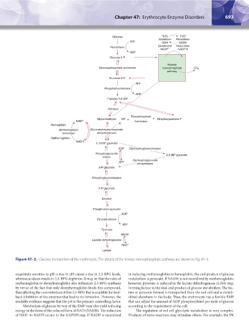
                  Figure 47–2.  Glucose metabolism of the erythrocyte. The details of the hexose monophosphate pathway are shown in Fig. 47–3.


                  exquisitely sensitive to pH: a rise in pH causes a rise in 2,3-BPG levels,   in reducing methemoglobin to hemoglobin, the end product of glucose
                  whereas acidosis results in 2,3-BPG depletion. It may be that the ratio of   metabolism is pyruvate. If NADH is not reoxidized by methemoglobin,
                  oxyhemoglobin to deoxyhemoglobin also influences 2,3-BPG synthesis   however, pyruvate is reduced in the lactate dehydrogenase (LDH) step,
                  by virtue of the fact that only deoxyhemoglobin binds this compound,   forming lactate as the final end product of glucose metabolism. The lac-
                  thus affecting the concentration of free 2,3-BPG that is available for feed-  tate or pyruvate formed is transported from the red cell and is metab-
                  back inhibition of the enzymes that lead to its formation. However, the   olized elsewhere in the body. Thus, the erythrocyte has a flexible EMP
                  available evidence suggests that the pH is the primary controlling factor.  that can adjust the amount of ADP phosphorylated per mole of glucose
                     Metabolism of glucose by way of the EMP may also yield reducing   according to the requirement of the cell.
                  energy in the form of the reduced form of NAD (NADH). The reduction   The regulation of red cell glycolytic metabolism is very complex.
                  of NAD  to NADH occurs in the GAPDH step. If NADH is reoxidized   Products of some reactions may stimulate others. For example, the PK
                        +


          Kaushansky_chapter 47_p0689-0724.indd   693                                                                   9/17/15   6:44 PM
   713   714   715   716   717   718   719   720   721   722   723