
Page 752 - Williams Hematology ( PDFDrive )
P. 752
726 Part VI: The Erythrocyte Chapter 48: The Thalassemias: Disorders of Globin Synthesis 727
CODON 6 - 1bp IVS 1 - 5 G→C
IVS 1 - 1G→A IVS 1 - 1 G→T
IVS 2 - 1G→A CODONS 41 - 42.4bp DEL
IVS 2 - 745C→G CODONS 26 GAG→AAG(HbE)
CODON 39 CAG→TAG
IVS 1 - 6T→C
IVS 1 -110 G→A
IVS 1 - 110 G→A IVS 2 - 654 C→T
IVS 1 - 5 G→C
IVS 1 - 6 T→C CODONS 41 - 42.4bp DEL.
CODON 17 AAG→TAG
CODON 39 CAG→TAG CODON 26 GAG→AAG(HbE)
CODON 8 2bp DEL
–28 A→G
–29A→G
IVS 1 - 5G→C
–29 A→G IVS 1 - 5 G→C
–88 C→T 619 bp DELETION
CODON 24 T→A CODON 8/9 + G
POLY-A T→C IVS 1 -1 G→T
CODONS 41 - 42.4bp DEL.
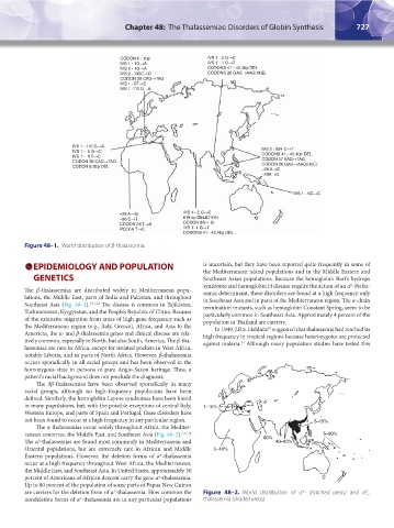
Figure 48–1. World distribution of β-thalassemia.
EPIDEMIOLOGY AND POPULATION is uncertain, but they have been reported quite frequently in some of
the Mediterranean island populations and in the Middle Eastern and
GENETICS Southeast Asian populations. Because the hemoglobin Bart’s hydrops
syndrome and hemoglobin H disease require the action of an α -thalas-
0
The β-thalassemias are distributed widely in Mediterranean popu- semia determinant, these disorders are found at a high frequency only
lations, the Middle East, parts of India and Pakistan, and throughout in Southeast Asia and in parts of the Mediterranean region. The α-chain
Southeast Asia (Fig. 48–1). 7,11,12 The disease is common in Tajikistan, termination mutants, such as hemoglobin Constant Spring, seem to be
Turkmenistan, Kyrgyzstan, and the People’s Republic of China. Because particularly common in Southeast Asia. Approximately 4 percent of the
of the extensive migration from areas of high gene frequency such as population in Thailand are carriers.
the Mediterranean region (e.g., Italy, Greece), Africa, and Asia to the In 1949, J.B.S. Haldane suggested that thalassemia had reached its
13
Americas, the α- and β-thalassemia genes and clinical disease are rela- high frequency in tropical regions because heterozygotes are protected
tively common, especially in North, but also South, America. The β-tha- against malaria. Although many population studies have tested this
13
lassemias are rare in Africa, except for isolated pockets in West Africa,
notably Liberia, and in parts of North Africa. However, β-thalassemia
occurs sporadically in all racial groups and has been observed in the
homozygous state in persons of pure Anglo-Saxon heritage. Thus, a
patient’s racial background does not preclude the diagnosis.
The δβ-thalassemias have been observed sporadically in many
racial groups, although no high-frequency populations have been
defined. Similarly, the hemoglobin Lepore syndromes have been found
in many populations, but, with the possible exceptions of central Italy, 1–15%
Western Europe, and parts of Spain and Portugal, these disorders have
not been found to occur at a high frequency in any particular region. 5–15%
The α-thalassemias occur widely throughout Africa, the Mediter-
ranean countries, the Middle East, and Southeast Asia (Fig. 48–2). 7,11,12 5–80%
The α -thalassemias are found most commonly in Mediterranean and 60% 40–80%
0
Oriental populations, but are extremely rare in African and Middle 5–40%
Eastern populations. However, the deletion forms of α -thalassemia
+
occur at a high frequency throughout West Africa, the Mediterranean,
the Middle East, and Southeast Asia. In United States, approximately 30
percent of Americans of African descent carry the gene α -thalassemia.
+
Up to 80 percent of the population of some parts of Papua New Guinea
are carriers for the deletion form of α -thalassemia. How common the Figure 48–2. World distribution of α - (hatched areas) and α _
+
0
+
nondeletion forms of α -thalassemia are in any particular populations thalassemia (shaded areas).
+
Kaushansky_chapter 48_p0725-0758.indd 727 9/18/15 2:57 PM