
Page 775 - Williams Hematology ( PDFDrive )
P. 775
750 Part VI: The Erythrocyte Chapter 48: The Thalassemias: Disorders of Globin Synthesis 751
Asia, is one of the most important hemoglobinopathies in the world
population. 7,9,10,234–240 As mentioned earlier in “Etiology and Pathogene-
sis,” hemoglobin E is synthesized at a reduced rate and hence produces
the clinical phenotype of a mild form of β-thalassemia. Hence, when
hemoglobin E is inherited with β-thalassemia—and most often this is
+
a β - or severe β -thalassemia mutation in Southeast Asia and India—a
0
marked deficit of β-chain production results, with the clinical picture
of severe β-thalassemia. Hemoglobin E thalassemia shows a remark-
able variability in clinical expression, 234–238 ranging from a mild form
of thalassemia intermedia to a transfusion-dependent condition clin-
ically indistinguishable from homozygous β-thalassemia. The reasons
for this variability of expression are not understood, although some of
the factors involved are identical to those that modify other forms of
β-thalassemia. 239,240
In more-severe cases of hemoglobin E thalassemia, severe anemia
with growth retardation, leg ulcers, bone deformity, marked tendency to
A infection, iron loading, and variable splenomegaly and hypersplenism
are seen. Large tumor masses composed of extramedullary erythropoi-
etic tissue may cause a variety of compression syndromes, including a
clinical picture that closely mimics a cerebral tumor. Another curious
picture that seems to be restricted to splenectomized patients is an oblit-
erative occlusion of the pulmonary vasculature that is believed to result
from an extremely high platelet count. 241
The clinical course and complications in transfusion-dependent
patients are similar to those observed in homozygous β-thalassemia.
In the milder forms, the main complications are progressive hyper-
splenism, organ damage as a result of progressive iron loading from
an increased rate of absorption, extramedullary erythropoietic tumor
masses, bone disease, and infection. The blood picture shows a typical
thalassemic pattern. The hemoglobin consists of E, F, and A . Usually
2
0
no hemoglobin A is present because the β -thalassemias are particularly
common in the parts of the world where hemoglobin E is found.
Newer studies emphasize the complex interactions between
B genetic factors, 239,240 differences in adaptation to anemia, particularly in
early life (see “Pathophysiology” above), and the environment, notably
proneness to malarial infection, that underlie the widely differing and
unstable phenotypes of patients with hemoglobin E β-thalassemia. 238,239
β-THALASSEMIA WITH NORMAL HEMOGLOBIN
A LEVEL
2
Rare forms of β-thalassemia are seen in which heterozygotes have nor-
mal hemoglobin A levels. Their main clinical importance is that they
2
can be confused with the more severe forms of α-thalassemia in the
heterozygous state and therefore may cause difficulties in genetic coun-
seling and prenatal diagnosis. Based on hematologic studies, two main
classes of “normal hemoglobin A β-thalassemia”—sometimes called
2
242
types 1 and 2—are seen. Type 1 is the “silent” form of β-thalassemia.
Type 2 is heterogeneous, with many cases representing the compound
C heterozygous state for β-thalassemia and δ-thalassemia.
“Silent” β-thalassemia 7,243 is characterized by no hematologic
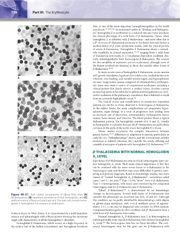
Figure 48–21. Acid elution preparations of blood films from (A) changes in heterozygotes. Several mild forms of β-thalassemia that
δβ-thalassemia, (B) hereditary persistence of fetal hemoglobin, and (C) underlie this phenotype are described (see Refs. 44 and 45). Although
artificial mixture of fetal and adult red cells. The dark cells contain hemo- this condition can be partly identified by demonstrating a mild degree
globin F. Hemoglobin F is resistant to acid elution. of globin-chain imbalance, with α-to-β synthesis ratios of approxi-
mately 1.5:1, it can only be diagnosed with certainty by DNA analysis.
0
Compound heterozygotes for this condition and β -thalassemia have a
it also is found in West Africa. It is characterized by a mild hemolytic mild form of β-thalassemia intermedia.
anemia and splenomegaly with a blood picture showing the numerous Normal hemoglobin A β-thalassemia type 2 in heterozygotes is
2
target cells characteristic of all the hemoglobin C disorders. indistinguishable from typical β-thalassemia with elevated hemoglobin
Hemoglobin E thalassemia, which occurs at a high frequency in A levels. The homozygous state has not been described. The com-
242
2
the eastern half of the Indian subcontinent and throughout Southeast pound heterozygous state for this gene and for β-thalassemia with
Kaushansky_chapter 48_p0725-0758.indd 750 9/18/15 2:58 PM