
Page 772 - Williams Hematology ( PDFDrive )
P. 772
746 Part VI: The Erythrocyte Chapter 48: The Thalassemias: Disorders of Globin Synthesis 747
3.5 to 7.0 percent. The level of fetal hemoglobin is elevated in approxi- α -Thalassemia and α -Thalassemia Traits
+
0
mately 50 percent of cases, usually to 1 to 3 percent and rarely to greater The α -thalassemia trait is characterized by the presence of 5 to 15 per-
0
than 5 percent. cent hemoglobin Bart’s at birth. This hemoglobin disappears during
7
maturation and is not replaced by a similar amount of hemoglobin H.
α-THALASSEMIAS An occasional cell with hemoglobin H inclusion bodies may appear
Hemoglobin Bart’s Hydrops Fetalis Syndrome after incubation with brilliant cresyl blue. This phenomenon is often
used as a diagnostic test for the α-thalassemia trait. However, the test
In infants with the hydrops fetalis syndrome, the blood film shows is difficult to standardize and requires much experience to be useful. In
severe thalassemic changes with many nucleated red cells. The hemo- adult life, the red cells of heterozygotes have morphologic changes of
globin consists mainly of hemoglobin Bart’s, with approximately 10 heterozygous thalassemia with low MCH and MCV values. The elec-
to 20 percent hemoglobin Portland. Usually no hemoglobin A or F is trophoretic pattern is normal. Globin-synthesis studies show a deficit
present, although rare cases that seem to result from interaction of α - of α-chain production, with an α-chain–to–β-chain production ratio of
0
thalassemia with a severe nondeletion form of α -thalassemia show approximately 0.7.
+
small amounts of hemoglobin A. The α -thalassemia trait (–α/αα) is characterized by a mild reduc-
+
tion in MCH and MCV values although in some cases there are normal
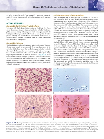
Hemoglobin H Disease values, 1 to 2 percent of hemoglobin Bart’s at birth in some but not all
The blood film shows hypochromia and anisopoikilocytosis. The retic- cases, and a slightly reduced α-chain–to–β-chain production ratio of
ulocyte count usually is approximately 5 percent. Incubation of the approximately 0.8; thus, this genotype often is referred to as silent car-
red cells with brilliant cresyl blue results in ragged inclusion bodies in rier. Extensive studies comparing the level of hemoglobin Bart’s at birth
almost all cells. These bodies form because of precipitation of hemoglo- with a DNA analyses demonstrated that there is no detectable hemoglo-
bin H in vitro as a result of redox action of the dye. After splenectomy, bin Bart’s in a significant number of newborns who are heterozygous for
large, single Heinz bodies are observed in some cells (Fig. 48–18). These α -thalassemia. 218,219 Globin gene synthetic ratios can be distinguished
+
bodies are formed by in vitro precipitation of the unstable hemoglobin from normal only by studying relatively large numbers of samples and
H molecule and are seen only after splenectomy. Hemoglobin H con- comparing the mean α–to–β ratio with that of normal control subjects.
stitutes between 5 and 40 percent of the total hemoglobin. Traces of This approach is not reliable for diagnosing individual cases of the
hemoglobin Bart’s may be present, and the hemoglobin A level usually α -thalassemia trait, and, unfortunately, no reliable method of diagnosis
+
2
is slightly subnormal. is available except for DNA analysis.
A B
C D
Figure 48–18. Hemoglobin H disease (α-thalassemia). Blood films. A. Note hypochromic red cells, anisocytosis, target cells, poikilocytes, includ-
ing tear-drop-shaped red cells. B. Wet preparation stained with crystal violet. Inclusions in red cells (Heinz bodies) usually attached to membrane.
C. Postsplenectomy. Note reduction in poikilocytes and frequency of target cells, a change consistent with hemoglobin H disease and enhanced by
postsplenectomy effects. A nucleated red cell is in this field, reflecting an increase in their prevalence in the blood after splenectomy. D. Blood incu-
bated for 90 minutes with brilliant cresyl blue. Numerous hemoglobin H intracellular precipitates (precipitates of excess β-globin chains). The frequent
crenation is an artifact of the incubation conditions. (Reproduced with permission from Lichtman's Atlas of Hematology, www.accessmedicine.com.)
Kaushansky_chapter 48_p0725-0758.indd 747 9/18/15 2:58 PM