
Page 771 - Williams Hematology ( PDFDrive )
P. 771
746 Part VI: The Erythrocyte Chapter 48: The Thalassemias: Disorders of Globin Synthesis 747
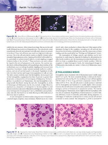
A B C
Figure 48–16. Blood films in β-thalassemia. A. β-Thalassemia minor. Anisocytosis, poikilocytosis, hypochromia. Occasional spherocytes and stoma-
tocytes. B. Scanning electron micrograph of cells in (A) showing more detail of the poikilocytes. Note the knizocyte (pinch-bottle cell) at the lower
right. C. β-Thalassemia major. Marked anisocytosis with many microcytes. Marked poikilocytosis. Anisochromia. Nucleated red cell on the right. Small
lymphocyte on the left. (Reproduced with permission from Lichtman’s Atlas of Hematology, www.accessmedicine.com.)
poikilocytes are common. After splenectomy, large, flat macrocytes and over β- and γ-chain production is always observed. Other aspects of the
small, deformed microcytes are frequently seen. The reticulocyte count laboratory findings in this condition, including red cell survival, iron
is moderately elevated, and nucleated red cells nearly always are present absorption, ferrokinetics, erythrokinetics, and the consequences of iron
in the blood. These red cell forms may reach very high levels after sple- loading, were discussed earlier (see “Etiology and Pathogenesis” above).
nectomy. The white cell and platelet counts are slightly elevated unless The examination of siblings, parents, and children can be very
secondary hypersplenism occurs. Staining of the blood with methyl vio- important in confirming the diagnosis by finding the abnormalities in
let, particularly in splenectomized subjects, reveals stippling or ragged other family members, and the examining physician should make every
169
inclusion bodies in the red cells. These inclusions can nearly always effort to obtain a complete blood count in family members. With the
be found in the red cell precursors in the marrow. The marrow usu- exception of higher hemoglobin levels, the hematological changes in
ally shows erythroid hyperplasia with morphologic abnormalities of the β-thalassemia intermedia are similar to those in β-thalassemia major
erythroblasts, such as striking basophilic stippling and increased iron (Fig. 48–17).
deposition. Iron kinetic studies indicate markedly ineffective erythro-
poiesis, and red cell survival usually is shortened. Populations of cells
with very short survival and longer-lived populations of cells are seen. β-THALASSEMIA MINOR
The latter contain relatively more fetal hemoglobin. An increased level Hemoglobin values of patients with β-thalassemia minor usually range
of fetal hemoglobin, ranging from less than 10 percent to greater than from 9 to 11 g/dL. The most consistent finding is small, poorly hemo-
90 percent, is characteristic of homozygous β-thalassemia. No hemo- globinized red cells (see Fig. 48–16), resulting in mean cell hemoglobin
0
globin A is produced in β -thalassemia. The fetal hemoglobin is het- (MCH) values of 20 to 22 pg and mean corpuscular volume (MCV)
erogeneously distributed among the red cells. Hemoglobin A levels values of 50 to 70 fL. The red cell count is usually normal or elevated
2
in homozygous β-thalassemia may be low, normal, or high. However, and the hemoglobin and hematocrit is usually slightly below normal;
expressed as a proportion of hemoglobin A, the hemoglobin A level however, the red cell indices are particularly useful in screening for het-
2
almost invariably is elevated. Differential centrifugation studies indicate erozygous carriers of thalassemia in population surveys. The marrow
some heterogeneity of hemoglobin F and A distribution among thalas- in heterozygous β-thalassemia shows slight erythroid hyperplasia with
2
semic red cells, but their level in whole blood gives little indication of rare red cell inclusions. Megaloblastic transformation as a result of folic
their total rates of synthesis. acid deficiency occurs occasionally, particularly during pregnancy. A
In vitro hemoglobin synthesis studies using marrow or blood show mild degree of ineffective erythropoiesis is noted, but red cell survival
a marked degree of globin-chain imbalance. Marked excess of α-chain is normal or nearly normal. The hemoglobin A level is increased to
2
A B
Figure 48–17. A. Thalassemia intermedia. Blood films. Marked anisocytosis, poikilocytosis with elliptical, oval, tear-drop-shaped, and fragmented
red cells. Target cells. B. Postsplenectomy. Morphology similar to that in (A) but with a nucleated red cell, coarsely stippled cell in center of field,
and large and numerous platelets, indicative of the changes superimposed by splenectomy. (Reproduced with permission from Lichtman's Atlas of
Hematology, www.accessmedicine.com.)
Kaushansky_chapter 48_p0725-0758.indd 746 9/18/15 2:58 PM