
Page 774 - Williams Hematology ( PDFDrive )
P. 774
748 Part VI: The Erythrocyte Chapter 48: The Thalassemias: Disorders of Globin Synthesis 749
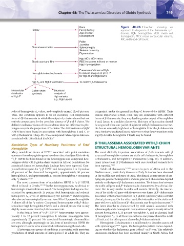
Figure 48–20. Flowchart showing an
approach to diagnosis of the thalassemia syn-
dromes. Hgb, hemoglobin; MCH, mean cell
hemoglobin; MCV, mean corpuscular volume;
RBC, red blood cell count.
reduced hemoglobin A values, and completely normal blood pictures. categorized under the general heading of heterocellular HPFH. Their
2
Thus, this condition appears to be an extremely well-compensated clinical importance is that, when they are coinherited with different
form of δβ-thalassemia in which the output of γ chains almost but not forms of β-thalassemia, they may lead to greater output of hemoglobin
entirely compensates for the complete absence of β and δ chains. The F and, hence, to a milder phenotype. This type of interaction should
different molecular forms of this condition show no difference in phe- be suspected when one parent of a patient with β-thalassemia interme-
notype except in the proportion of γ chains. The African forms of (δβ) dia has an unusually high level of hemoglobin F for the β-thalassemia
G
0
HPFH have been found in association with hemoglobins S and C or trait. Similarly, unaffected lateral relatives or other family members with
with β-thalassemia (Chap. 49). These compound heterozygous states are slightly elevated hemoglobin F levels may be found.
associated with little clinical disability. 7
β-THALASSEMIA ASSOCIATED WITH β-CHAIN
Nondeletion Types of Hereditary Persistence of Fetal
Hemoglobin STRUCTURAL HEMOGLOBIN VARIANTS
Many nondeletion forms of HPFH associated with point mutations The most clinically important associations of β-thalassemia with β
upstream from the γ-globin genes have been described (see Table 48–4). structural hemoglobin variants are sickle cell thalassemia, hemoglobin
+
G γ β HPFH has been found in the heterozygous and compound hete- C thalassemia, and hemoglobin E thalassemia (Chap. 49). In addition,
rozygous states with β-globin chain variants in African populations. No many interactions of β-thalassemia with rare structural variants have
associated clinical or hematologic findings have been reported. Com- been reported. 7,9,10
pound heterozygotes for γ β HPFH and hemoglobins S or C produce Sickle cell thalassemia 7,232,233 occurs in parts of Africa and in the
G
+
45 percent of the abnormal hemoglobin, approximately 30 percent Mediterranean, particularly Greece and Italy. It also has been observed
hemoglobin A, and approximately 20 percent hemoglobin F containing in the Middle East and parts of India. The clinical consequences of car-
only γ chains. 225,226 rying one gene for hemoglobin S and one gene for β-thalassemia depend
G
The most common form of nondeletion HPFH is γ β HPFH, entirely on the type of β-thalassemia mutation. The interaction between
+
A
which is found in Greeks. 227–229 In the homozygous state, no clinical or the sickle cell gene and β -thalassemia is characterized by a clinical dis-
0
hematologic abnormalities are noted. The hemoglobin findings are char- order that is very similar to sickle cell anemia. Similarly, the interac-
acterized by approximately 25 percent fetal hemoglobin and reduced tion of the sickle cell gene with the more severe forms of β -thalassemia
+
hemoglobin A levels of approximately 0.8 percent. Heterozygotes, associated with marked reduction in β-globin synthesis yields a similar
230
2
who also are hematologically normal, have 10 to 15 percent hemoglobin clinical phenotype. On the other hand, the interaction of the sickle cell
F, almost all of the γ variety. Compound heterozygotes with β-thalas- gene with very mild forms of β -thalassemia may be quite innocuous.
A
233
+
semia have high hemoglobin F levels and a clinical picture that is only The latter disorder is characterized by mild anemia associated with
slightly more severe than the β-thalassemia trait. splenomegaly and a hemoglobin composition of approximately 60 to 70
In the British form of γ β HPFH heterozygotes have approx- percent hemoglobin S, 25 percent hemoglobin A, and an elevated level
A
231
+
imately 5 to 12 percent hemoglobin F, whereas homozygotes have of hemoglobin A . In all these interactions, one parent shows the sickle
2
approximately 20 percent. No associated hematologic abnormalities cell trait, and the other parent shows the β-thalassemia trait.
are seen, although surprisingly in this form of nondeletion HPFH the Hemoglobin C thalassemia is a mild hemolytic disorder associated
hemoglobin F seems to be unevenly distributed among the red cells. with splenomegaly. 7,9,10 Again, the hemoglobin pattern varies depend-
A heterogeneous group of conditions is associated with persistent ing on whether the thalassemia gene is the β or β type. This relatively
0
+
production of small amounts of hemoglobin F in adult life. They are innocuous condition has been recorded mainly in North Africa, but
Kaushansky_chapter 48_p0725-0758.indd 749 9/18/15 2:58 PM