
Page 773 - Williams Hematology ( PDFDrive )
P. 773
748 Part VI: The Erythrocyte Chapter 48: The Thalassemias: Disorders of Globin Synthesis 749
Homozygous State for Nondeletion Types of α-Thalassemia hemoglobin F levels encountered in juvenile chronic myelogenous leu-
The homozygous state for nondeletion forms of α-thalassemia involv- kemia, this disorder may superficially resemble β-thalassemia. How-
ing the dominant (α ) globin gene causes a more severe deficit of ever, the finding of primitive cells in the marrow, the absence of elevated
2
α chains than do the deletion forms of α -thalassemia. In some cases, hemoglobin A levels on hemoglobin electrophoresis, the decrease in
+
2
the homozygous state produces hemoglobin H disease. carbonic anhydrase in juvenile chronic myelogenous leukemia, and
The homozygous state for hemoglobin Constant Spring or other characteristic in vitro responses of myeloid progenitors in vitro to gran-
chain-termination mutations is associated with moderately severe ulocyte-monocyte colony-stimulating factor (Chap. 87) readily differ-
hemolytic anemia in which, for reasons not explained, no hemoglo- entiate this disorder from β-thalassemia.
bin H is present but small amounts of hemoglobin Bart’s persist into
adult life. The homozygous states for the other nondeletion forms of α - LESS-COMMON FORMS OF THALASSEMIA
+
thalassemia are associated with hemoglobin H disease. In the homozy-
0
gous state for hemoglobin Constant Spring, the blood picture shows (δβ) -Thalassemia
mild thalassemic changes with normal-size red cells. 216,217 The hemo- The homozygous state for δβ-thalassemia is clinically milder than
globin consists of approximately 5 to 6 percent hemoglobin Constant Cooley anemia and is one form of thalassemia intermedia. 220–222 Only
Spring, normal hemoglobin A levels, and trace amounts of hemoglobin hemoglobin F is present; hemoglobins A and A are not produced. Het-
2
2
Bart’s. The remainder is hemoglobin A. erozygous δβ-thalassemia is hematologically similar to β-thalassemia
7
The heterozygous state for hemoglobin Constant Spring shows no minor. The fetal hemoglobin level is higher (range: 5 to 20 percent),
hematologic abnormality. The hemoglobin pattern is normal except for and the hemoglobin A value is normal or slightly reduced. As in β-
2
the presence of approximately 0.5 percent hemoglobin Constant Spring. thalassemia, the fetal hemoglobin is heterogeneously distributed among
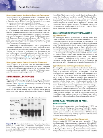
The latter can be observed on alkaline starch-gel electrophoresis as a the red cells, thus distinguishing this disorder from HPFH (Fig. 48–21).
faint band migrating between hemoglobin A and the origin. It is best Heterozygosity for both β-thalassemia and δβ-thalassemia results
2
seen on heavily loaded starch gels and is easily missed if other electro- is a condition clinically similar to but milder than Cooley anemia. The
phoretic techniques are used (Fig. 48–19). In the newborn, usually 1 to hemoglobin consists largely of hemoglobin F, with a small amount
3 percent hemoglobin Bart’s is present in the cord blood. of hemoglobin A . This finding is seen because the associated β-
2
thalassemia gene has usually been the β variety. δβ-Thalassemia has
0
+
Homozygous State for Deletion Forms of α -Thalassemia also been observed in individuals heterozygous for hemoglobin S or C. 7
The homozygous state for deletion forms of α -thalassemia is charac-
+
+
terized by a thalassemic blood picture with 5 to 10 percent hemoglobin (δβ) -Thalassemia and Hemoglobin Lepore Disorders
Bart’s at birth and hematologic findings similar to those in α -thalas- The hemoglobin Lepore disorders have been described in the homozy-
0
semia heterozygotes in adult life. In general, the –α deletion is associ- gous state and in the heterozygous state, either alone or in association
4.2
ated with a more severe phenotype than is the –α deletion. 7 with β- or δβ-thalassemia, hemoglobin S, or hemoglobin C. 7,9,223 In the
3.7
homozygous state, approximately 20 percent of the hemoglobin is of
DIFFERENTIAL DIAGNOSIS the Lepore type and 80 percent is fetal hemoglobin. Hemoglobins A
and A are absent. The clinical picture is variable. Some cases are iden-
2
The clinical and hematologic findings in homozygous β-thalassemia tical to transfusion-dependent homozygous β-thalassemia; others are
and hemoglobin H disease are so characteristic that the diagnosis usu- associated with the clinical picture of thalassemia intermedia. In the
ally is not difficult. Figure 48–20 shows a simple flowchart for laboratory heterozygous state, the findings are similar to those of β-thalassemia
investigations of a suspected case. minor. The hemoglobin consists of approximately 10 percent hemoglo-
In early childhood, distinguishing the thalassemias from the bin Lepore, with a reduced level of hemoglobin A and a slight but con-
2
congenital sideroblastic anemias may be difficult, but the marrow sistent increase in fetal hemoglobin level. The Lepore hemoglobins have
appearances in the latter are quite characteristic. Because of the high been found sporadically in most racial groups. In the majority of cases,
chemical analysis has shown that these hemoglobins are identical to
hemoglobin Lepore Washington-Boston. Hemoglobin Lepore Hollan-
dia and Lepore Baltimore have been observed in only a few patients. 7,223
1 2 3 4 5 6
Origin
Hgb Constant HEREDITARY PERSISTENCE OF FETAL
Spring HEMOGLOBIN
Hgb A 2
The current knowledge about the molecular pathology of HPFH was
described earlier in “Etiology and Pathogenesis.” Table 48–4 summa-
Hgb A rizes the currently accepted classification and nomenclature of this
complex group of conditions. The different forms of HPFH are of very
Hgb Bart’s little clinical importance except that they may interact with thalassemia
or the structural hemoglobin variants.
Hgb H (δβ) Hereditary Persistence of Fetal Hemoglobin
0
+
Homozygotes for (δβ) HPFH have 100 percent hemoglobin F. Their
0
blood shows mild thalassemic changes, with reduced MCH and MCV
Figure 48–19. Hemoglobin (Hgb) Constant Spring. Starch gel electro-
phoresis of 1,2, normal adult; 3,4, compound heterozygotes for hemo- values very similar to those observed in heterozygous β-thalassemia.
globin Constant Spring and α -thalassemia with hemoglobin H disease; Similarly, they have imbalanced globin chain production, with ratios
0
224
0
5, normal adult; and 6, compound heterozygote for α -thalassemia and in the range of those observed in β-thalassemia heterozygotes. Het-
hemoglobin Constant Spring. erozygotes have approximately 20 to 30 percent hemoglobin F, slightly
Kaushansky_chapter 48_p0725-0758.indd 748 9/18/15 2:58 PM