
Page 78 - Williams Hematology ( PDFDrive )
P. 78
54 Part II: The Organization of the Lymphohematopoietic Tissues Chapter 5: Structure of the Marrow and the Hematopoietic Microenvironment 55
32
Prenatal Postnatal display a specific integrin (Mac-1) that is not found in marrow HSCs.
Cellularity In the last third of gestation, the HSCs and early hematopoietic pro-
(%)
genitor cells migrate from the fetal liver through the circulation seed-
100 Bone
Yolk sac ing the spleen and marrow. Fetal liver hematopoiesis declines steadily
marrow
80 Liver Vertebra as the spleen and marrow become the major hematopoietic sites. At
birth, the marrow is the major hematopoietic site in humans, while the
60 Sternum spleen remains a prominent but decreasing site in the mouse (Chap. 7).
Visceral endoderm is in close proximity to the mesoderm formed
40
Spleen Tibia Rib by gastrulation in those sites where HSCs are generated in the embryo.
20 Femur This proximity is important in that the endoderm appears to induce
both endothelial and blood cell development in the adjacent mesoderm
0
12 34 56 78 9 10 20 30 40 50 60 70 through secretion of Indian hedgehog (IHH), a member of the hedge-
Birth
33
Fetal months Age in years hog family of proteins. IHH, in turn, upregulates the expression of
BMP4 in the developing mesodermal cells. BMP4 upregulation is
33
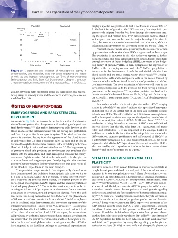
Figure 5–1. Expansion and recession of hematopoietic activity in important for the development of both the endothelial cells that form
extramedullary and medullary sites. For details regarding the nature blood vessels and the HSCs located within these vessels. 33,34 Develop-
of yolk sac and hepatic hematopoiesis, see “Sites of Hematopoiesis: ing endothelial cells and hematopoietic cells in the vessels formed by
Embryogenesis and Early Stem Cell Development.” Chapter 7 provides these endothelial cells are found in each site of primitive and defini-
a more comprehensive treatment of this topic (see Fig. 7–1 in Chap. 7).
tive hematopoiesis. The close association of these two cell types in the
developing embryo has led to the proposal for their having a common
using in vitro long-term progenitor assays and surrogate in vivo repopu- precursor, the hemangioblast. 35,36 Important proteins involved in the
lating assays in severely immunodeficient mice and xenogeneic animal development of the hemangioblasts are BMP4, VE growth factor recep-
models (Chap. 18). tor KDR/Flk-1, transcription factor TAL1, and TAL1’s binding partner
LMO2. 35,36
37
Marked endothelial cells in mice give rise to the HSCs. Imaging
SITES OF HEMATOPOIESIS studies in zebrafish 38,39 and mice indicate that specialized hemogenic
40
EMBRYOGENESIS AND EARLY STEM CELL endothelial cells in the ventral part of the aorta can transform without
mitosis into HSCs. The differentiation of HSCs from hemangioblasts
DEVELOPMENT and/or hemogenic endothelium requires the signaling protein Notch1
As shown in Fig. 5–1, the marrow is the last in a series of anatomical and the transcription factors GATA-2, MYB, and Runx1. 35,36,41,42 The
sites of hematopoiesis that change several times during embryonic and mechanism driving this earliest expansion of HSC is not well-defined,
fetal development. 25–28 The earliest hematopoietic cells develop in the but two factors that also play roles later, KIT ligand/stem cell factor
blood islands of the extraembryonic yolk sac during late gastrulation (SCF) and interleukin (IL)-3, are important in the embryo. BMP4, in
and form the primitive hematopoietic system. This primitive hemato- addition to its role in the induction of hematopoietic and endothelial
poiesis is transient, lasting from the appearance of the blood islands differentiation, increases proliferative and self-renewal of HSCs 33,34 as
at embryonic days 7.5 days postcoitum (dpc) in mice and 19 dpc in it differentially upregulates KIT (SCF receptor) in the HSCs, but not in
43
humans through the final cellular divisions in the circulating embryonic adjacent endothelial cells. Expansion of the earliest definitive HSC is
blood at 13.5 dpc in mice and week 6 in humans. 28,29 The large majority also mediated by Notch signaling as it induces the Runx1 transcription
of primitive blood cells produced are erythrocytes that enucleate after factor 41,42 and one of its targets, the IL-3 gene. 44
release into the circulation, and their hemoglobin contains the embry-
onic α- and β-globin chains. Primitive hematopoietic cells also give rise STEM CELL AND MESENCHYMAL CELL
to macrophages and megakaryocytes. Overlapping with this transient
primitive hematopoiesis is definitive hematopoiesis that gives rise to all PLASTICITY
of the blood cell types found in the adult (Chap. 7). Primitive stem cells from human fetal liver or marrow reconstitute all
Transplantation experiments in hematopoietically ablated mice lymphohematopoietic-derived cells and part of the stromal microenvi-
have demonstrated that definitive hematopoietic cells arise on 8.5 to ronment in in vivo repopulation assays. These observations are con-
45
11.5 dpc in mice and weeks 4 to 6 in humans in three different embry- sistent with the early derivation of hematopoietic, vascular, and stromal
onic locations: the yolk sac blood islands, the anterior portion of the cells from a CD34−, KDR/Flk-1+, multipotential mesenchymal stem
47
aorta-gonad-mesonephros (AGM) region, and the allantoic portion of cell. 14–16,46 Identification of AC133+, CD34−, CD7− HSCs and demon-
48
the developing placenta. 26–28 The definitive murine erythroid cells cir- stration of endothelial precursors in AC133+ progenitor cells under-
culating on 8.5 to 11.5 dpc appear to be descendent from a transient score the crosstalk between hematopoiesis and angiogenesis signaling
population of erythroid/myeloid progenitors derived from the yolk pathways and establish the functional role of hemangioblasts in ontog-
sac, rather than being derived from HSCs that arise in the placenta and eny. 49–51 As early fetal hematopoiesis is established, the yolk sac vascular
AGM as occurs at later times in the fetus and adult. Serial transplanta- networks remain active sites of progenitor production and hemato-
30
28
tion in irradiated mice demonstrated that the earliest appearance of the poiesis. Long-term reconstituting HSCs express two members of the
intraembryonic human HSCs is in the AGM at week 5. HSCs migrate ATP-binding cassette genes (ABCG-2 and P-glycoprotein), allowing
31
through the blood to the fetal liver where they seed and mature into all the efflux of mitochondrial vital dyes such as Hoechst 33342 and rho-
of the cellular elements of the blood. 25–28 Erythrocytes, the predominant damine 123 and their isolation by multiparameter flow cytometry based
cell produced by definitive hematopoiesis during prenatal development, on their low side scatter (side population [SP] cells). 21–24 Enrichment of
52
are smaller than the primitive erythrocytes, and their hemoglobin con- the SP population for HSC has been achieved in both adult marrow
tains the fetal and adult globin chains. In mid-gestation, the HSCs that and fetal liver populations by using the signaling lymphocyte and
53
have migrated to the fetal liver undergo an exponential expansion and activation markers (SLAMs) to select cells with the specific phenotype
Kaushansky_chapter 05_p0051-0084.indd 54 9/19/15 12:10 AM