Page 82 - Williams Hematology ( PDFDrive )
P. 82

58             Part II:  The Organization of the Lymphohematopoietic Tissues                                                              Chapter 5:  Structure of the Marrow and the Hematopoietic Microenvironment                59


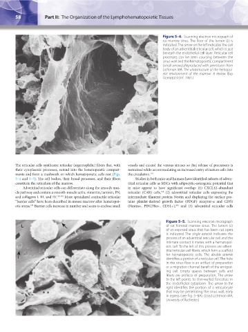
                                                                                       Figure 5–4.  Scanning electron micrograph of
                                                                                       rat marrow sinus. The floor of the lumen (L) is
                                                                                       indicated. The arrow on the left indicates the cell
                                                                                       body of an adventitial reticular cell, which is just
                                                                                       beneath the endothelial cell layer. Reticular cell
                                                                                       processes can be seen coursing between the
                                                                                       sinus wall and the hematopoietic compartment
                                                                                       (small arrows).(Reproduced with permission from
                                                                                       Lichtman MA: The ultrastructure of the hemopoi-
                                                                                       etic environment of the marrow: A review.  Exp
                                                                                       Hematol 9:391, 1981.)


                                                    L


               The reticular cells synthesize reticular (argentophilic) fibers that, with   vessels and extend the venous sinuses so that release of precursors is
               their cytoplasmic processes, extend into the hematopoietic compart-  restrained while accommodating an increased entry of mature cells into
               ments and form a meshwork on which hematopoietic cells rest (Figs.   the circulation. 131
               5–4 and 5–5). The cell bodies, their broad processes, and their fibers   Studies in both mice and humans have identified subsets of adven-
               constitute the reticulum of the marrow.                titial reticular cells as MSCs with adipocytic-osteogenic potential that
                   Adventitial reticular cells can differentiate along the smooth mus-  in mice appear to have significant overlap: (1) CXCL12–abundant
               cle pathway and contain α smooth-muscle actin, vimentin, laminin, FN,   reticular (CAR) cells,  (2) adventitial reticular cells expressing the
                                                                                      132
               and collagens I, III, and IV. 129,130  More specialized contractile reticular   intermediate filament protein Nestin and displaying the surface pro-
               “barrier cells” have been described in mouse marrow after hematopoi-  teins platelet-derived growth factor (PDGF) receptor-α and CD51
               etic stress.  Barrier cells increase in number and seem to enclose small   (Nestin+, PDGFRα+, CD51+),  and (3) adventitial reticular cells
                                                                                             133
                       131

                                                                                         Figure 5–5.  Scanning electron micrograph
                  L                                                                      of rat femoral marrow sinus. The lumen (L)
                                                                                         of an exposed sinus that has been cut open
                                                                                         is  indicated. The  single asterisk indicates the
                                                                                         process of an adventitial reticular cell and the
                                                                                         intimate contact it makes with a hematopoi-
                                                                                         etic cell. To the left of this process are adven-
                                                                                         titial reticular cell fibers, which form a scaffold
                                                           ∗                             for hematopoietic  cells.  The  double asterisk
                                                                                         identifies a portion of a reticular cell. The hole
                                                                                         in the sinus floor is an artifact of preparation
                                                                                         or a migration channel bereft of the emigrat-
                                                                                         ing cell. Empty spaces between cells and
                                                                                         fibers are artifacts of preparation. The arrow
                                                                                         to the left points to thin-walled fenestrae in
                                                                                         the endothelial cytoplasm. The  arrow to the
                                                                                         right identifies the portion of a reticulocyte
                             ∗ ∗                                                         that may be penetrating the sinus wall, early
                                                                                         in egress (see Fig. 5–8A). (Used Lichtman MA,
                                                                                         University of Rochester.)


          Kaushansky_chapter 05_p0051-0084.indd   58                                                                    9/19/15   12:10 AM
   77   78   79   80   81   82   83   84   85   86   87