
Page 83 - Williams Hematology ( PDFDrive )
P. 83
58 Part II: The Organization of the Lymphohematopoietic Tissues Chapter 5: Structure of the Marrow and the Hematopoietic Microenvironment 59
expressing leptin receptors. 115,134 The human equivalents are a popu- Adipocytes
lation of CD45−, CD146+ adventitial reticular cells that have smaller Adipocytes in the marrow develop by lipogenesis in fibroblast-like cells
subsets that express Nestin, PDGFRα, and CD51. A major proportion (Fig. 5–6). Reticular cells in mouse and human marrow can undergo
133
of these subsets are restricted to the perivascular area, but have some transformation to fat cells in vitro and can revert into fibroblasts in
cells scattered throughout the hematopoietic marrow. However, because culture by lipolysis, 101,143 and the Nestin+ MSCs and CAR MSCs can
138
these adventitial reticular subsets are also the major sources of CXCL12 differentiate into adipocytes. A reciprocal relationship between adipo-
and SCF in the marrow, they have important roles in establishing the cyte and osteoblast differentiation of MSCs appears to be controlled by
HSC niche near the marrow microvasculature while their progeny multiple transcription factors, with both peroxisome proliferator-acti-
establish the endosteum and its associated hematopoietic niche in the vated receptor-γ2 (PPARγ2) and CCAAT/enhancer binding protein
144
marrow. (C/EBP) promoting adipocyte differentiation. Marrow fat cells are
145
The majority of CAR cells are in close association with the sinus- relatively resistant to lipolysis during starvation, and their phenotype is
oidal endothelial cells but some are also associated with the endosteum. consistent with both white and brown fat. 146,147 Although the proportion
Like the Nestin+ MSCs, CAR cells appear to be progenitors of osteoblasts of saturated fatty acids is lower than in other fat deposits, marrow fat
135
and adipocytes while producing major amounts of CXCL12 and SCF. composition depends on whether it is located in the red, hematopoiet-
Development of CAR cells and their production of CXCL12 and SCF ically active, or the yellow, hematopoietically inactive, marrow. Human
136
is associated with the expression of the transcription factor Fox1c. marrow adipocytes support the differentiation of late-stage, commit-
CAR cells and the niches that they create in the marrow are required ted, myeloid and lymphoid hematopoietic cells, but they are unable
for the normal development of HSC, various differentiation stages of B- to support earlier progenitor stages. Quantification of immature
148
lymphocytes, natural killer cells, and the plasmacytoid dendritic cells hematopoietic cells including HSCs shows reduced numbers in human
that are all found in close physical association with CAR cells. 137 marrows with increased fat, and in vivo studies in mice confirm that
70
Autonomic neurons innervate the perivascular Nestin+, PDG- marrow adipocytes create a negative hematopoietic microenvironment
FRα+, CD51+ adventitial reticular cells which maintain the HSC niche that reduces development of HSCs and early-stage common hemato-
by several surface-displayed and/or secreted products including IL-7 poietic progenitors. 72
138
and VCAM-1, in addition to SCF and CXCL12. β-Adrenergic neuro-
transmission inhibits the expression of these proteins so that mice with Bone Cells
specific denervation have decreased marrow cellularity and increased Osteoblasts, osteoclasts, and elongated flat cells with a spindle-shaped
139
circulating hematopoietic progenitors. The sympathetic nervous sys- nucleus form the marrow endosteal lining. These endosteal cells and
149
tem controls circadian fluctuations in circulating HSC numbers though the closely associated microvascular cells participate in a dynamic pro-
its effect on MSC expression of the chemokine CXCL12 in the mar- cess in which endochondral bone formation proceeds with removal of
140
row. Studies in mice with defective myelinization and mice treated calcified cartilage and connective tissues by macrophages while new
with adrenergic antagonists or agonists indicate that the adrenergic bone is formed by osteoblasts and remodeled by specialized osteo-
nervous system in the marrow also regulates mobilization of HSCs by clasts. 114,150 Osteoblasts that become embedded in the bone matrix
granulocyte colony-stimulating factor (G-CSF). However, the non- proteins are termed osteocytes, a terminally differentiated cell that has
141
myelinating Schwann cells associated with the autonomic nerves of secretory capacity and influences the activities of osteoblasts, osteo-
the marrow secrete transforming growth factor-β (TGF-β) and thereby clasts, and hematopoietic cells. Resting endosteal cells express vimentin,
maintain the HSC quiescence. 142 tenascin, α smooth-muscle actin, osteocalcin, CD51, and CD56. They
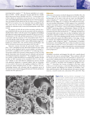
Figure 5–6. Scanning electron micrograph of rat
femoral marrow. Several sinuses and the intervening
hematopoietic cords are evident. The exposed lumen
(L) of one branching sinus is indicated. The sinus,
just above the L, contains a bean-shaped proplatelet
with an attenuated strand connected to a separat-
ing smaller proplatelet fragment. Smaller proplatelet
fragments are below the L. The short horizontal arrow
points to the cytoplasm of a transected megakaryo-
cyte. The lower arrow points to a fat cell. The rat femoral
marrow contains a modest number of fat cells. Spaces
in the hematopoietic cords are artifacts resulting from
transecting the femur. (Reproduced with permission
from Lichtman MA: The ultrastructure of the hemopoi-
etic environment of the marrow: A review. Exp Hematol
9:391, 1981.)
L
Kaushansky_chapter 05_p0051-0084.indd 59 9/19/15 12:10 AM