
Page 786 - Williams Hematology ( PDFDrive )
P. 786
760 Part VI: The Erythrocyte Chapter 49: Disorders of Hemoglobin Structure: Sickle Cell Anemia and Related Abnormalities 761
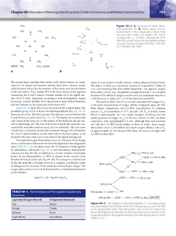
CH 2 Leu Figure 49–2. A. Structure of heme (ferro-
F4 (83) protoporphyrin IX). B. Heme group and its
CH H CH 3 environment in the unliganded α-chain. Only
C Leu H19 selected side chains are shown; the heme
H C CH CH 2 Leu F7 (86) (136) 4-propionate is omitted. (Reproduced from
3
N N His C e 1 Lys Gelin BR, Lee AW, Karplus M: Hemoglobin tertiary
F8 (87)
N e 2 E10 (61) E11 (62) structural change on ligand binding. J Mol Biol
Val
HC Fe CH Leu C δ 25;171(4):489–559, 1983.)
FG3 (91)
4 His
N
E7 (58)
N
C Val FG5 (93) 3 Leu G8
H 3
CH 3 (101)
C Phe
CD4
CH 2 H CH 2 (46)
Tyr y
HOOC · CH 2 CH · COOH C7 (42) x
2
z
A B
Hb nomenclature specifies that amino acids within helices are desig- where K is an empiric overall constant without physicochemical basis.
nated by the amino acid number and the helix letter, whereas amino The slope n is taken as a convenient measure of cooperativity. Values of
acids between helices bear the number of the amino acid and the letters n in noninteracting Hbs that exhibit hyperbolic, not sigmoid, oxygen
of the two helices. Thus, residue EF3 is the third residue of the segment dissociation curves (e.g., myoglobin) are approximately 1. In a normal
connecting the E and F helices, whereas residue F8 is the eighth resi- tetrameric Hb with four oxygen-reactive sites, the maximum value for n
due of the F helix. Alignment according to helical designation makes is 4.0; however, n values of 2.7 to 3.0 are found in normal Hb.
homology evident: Residue F8 is the proximal heme linked histidine, The point at which the Hb is one-half saturated with oxygen (P )
50
and the histidine on the distal side of the heme is E7. is the usual measurement of oxygen affinity. It depends upon pH (the
Figure 49–1B show the tertiary structure of the α and β chains. The Bohr effect), temperature, and 2,3-BPG concentration. In common
prosthetic group of Hb is heme (ferroprotoporphyrin IX); Fig. 49–2A practice, P is standardized at 37°C and pH 7.20. P of freshly drawn
50
50
shows its structure. The heme group is located in a crevice between the blood is approximately 26.7 torr under standard conditions, but the
E and F helices in each chain (Fig. 49–2B). The highly polar propionate partial pressure of oxygen (P ) of Hb from which 2,3-BPG has been
O2
side chains of the heme are on the surface of the molecule and are ion- removed is only approximately 13 torr. Although fetal and newborn
ized at physiologic pH. The rest of the heme is inside the molecule, sur- red cells have 2,3-BPG levels similar to those of adults, their oxygen
rounded by nonpolar residues except for two histidines. The iron atom dissociation curve is left shifted (increased oxygen affinity) with a P
50
is linked by a coordinate bond to the imidazole nitrogen (N) of histidine of approximately 23 torr because HbF does not react as strongly with
F8. The E7 distal histidine, on the other side of the heme plane, is not 2,3-BPG as does HbA.
bonded to the iron atom, but is very close to the ligand-binding site.
The sigmoid oxygen dissociation curve is a function of the change
of the conformation of the molecule from the liganded to the unliganded
state (Table 49–1). In the deoxy state, the Hb tetramer is held together
by intersubunit salt bonds (Fig. 49–3) and intersubunit hydrophobic
contacts (see Fig. 49–1B), in addition to a certain number of hydrogen
bonds. In deoxyhemoglobin, 2,3-BPG is situated in the central cavity
between the two β chains (see Fig. 49–1B). The change in conformation
of the Hb molecule is brought about by a complex, coordinated series
of changes in the structure of the molecule as heme binds oxygen. The
oxygen dissociation curve can be linearized by a transformation known
as the Hill plot:
log[y/(1 – y)] = log K+n log P
O2
TABLE 49–1. Nomenclature of Hemoglobin Quaternary
Structures
Liganded (Oxygen Bound) Unliganded (Reduced)
Oxy Deoxy Figure 49–3. Salt bridges in deoxyhemoglobin (* = ionizable group
less protonated at pH 9.0 than at pH 7.0). These groups account for 60%
R-state T-state of the alkaline Bohr effect. The remainder is due to αH5 His. (Data from
Relaxed Tense Perutz MF, Wilkinson AJ, Paoli M, et al: The stereochemical mechanism of the
cooperative effects in hemoglobin revisited, Annu Rev Biophys Biomol Struct
High affinity Low affinity 1998;27:1-34.)
Kaushansky_chapter 49_p0759-0788.indd 761 9/18/15 3:01 PM