
Page 789 - Williams Hematology ( PDFDrive )
P. 789
764 Part VI: The Erythrocyte Chapter 49: Disorders of Hemoglobin Structure: Sickle Cell Anemia and Related Abnormalities 765
+
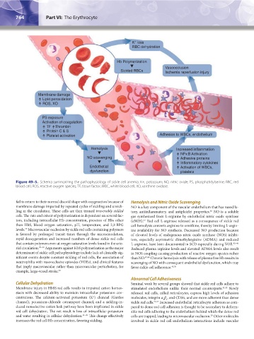
K loss
RBC dehydration
Hb Polymerization
Vasoocclusion
Sickled RBCs Ischemia reperfusion injury
Membrane damage
Lipid peroxidation
ROS, XO
PS exposure
Activation of coagulation
TF Thrombin
Protein C & S
Platelet activation Adhesion to WBCs, endothelium
Hemolysis Increased inflammation
NFκ B Activation
NO scavenging Adhesive proteins
Inflammatory cytokines
Endothetial Activation of WBCs,
dysfunction platelets
Figure 49–5. Schema summarizing the pathophysiology of sickle cell anemia. K+, potassium; NO, nitric oxide; PS, phosphatidylserine; RBC, red
blood cell; ROS, reactive oxygen species; TF, tissue factor; WBC, white blood cell; XO, xanthine oxidase.
fail to return to their normal discoid shape with oxygenation because of Hemolysis and Nitric Oxide Scavenging
membrane damage imparted by repeated cycles of sickling and unsick- NO is a key component of the vascular endothelium that has vasodila-
ling in the circulation. These cells are then termed irreversibly sickled tory, antiinflammatory, and antiplatelet properties. NO is a soluble
40
cells. The rate and extent of polymerization is dependent on several fac- gas synthesized from L-arginine by endothelial nitric oxide synthase
tors, including intracellular Hb concentration, presence of Hbs other (eNOS). Red cell L-arginase released as a consequence of sickle red
41
than HbS, blood oxygen saturation, pH, temperature, and 2,3-BPG cell hemolysis converts arginine to ornithine, thereby limiting L-argin-
levels. Microvascular occlusion by sickle red cells containing polymers ine availability for NO synthesis. Decreased NO production because
29
is favored by prolonged transit times through the microcirculation, of elevated levels of endogenous nitric oxide synthase (NOS) inhibi-
rapid deoxygenation and increased numbers of dense sickle red cells tors, especially asymmetric dimethylarginine (ADMA) and reduced
that contain polymers even at oxygen saturation levels found in the arte- L-arginine, have been documented in SCD especially during VOE. 42–46
rial circulation. 29–32 Arguments against HbS polymerization as the major Reduced plasma arginine levels and elevated ADMA levels also result
determinant of sickle cell pathophysiology include lack of clinically sig- in NOS coupling causing production of reactive oxygen species rather
nificant events despite constant sickling of red cells, the association of than NO. 47,48 Chronic hemolysis with release of plasma free Hb results in
neutrophilia with vasoocclusive episodes (VOEs), and clinical features scavenging of NO with consequent endothelial dysfunction, which may
that imply macrovascular rather than microvascular perturbation, for favor sickle cell adherence. 49,50
example, large-vessel stroke. 33
Abnormal Cell Adhesiveness
Cellular Dehydration Seminal work by several groups showed that sickle red cells adhere to
Membrane injury in HbSS red cells results in impaired cation homeo- stimulated endothelium unlike their normal counterparts. 51,52 Newly
stasis with decreased ability to maintain intracellular potassium con- released red cells, called reticulocytes, express high levels of adhesion
centrations. The calcium-activated potassium (K ) channel (Gardos molecules, integrin α β , and CD36, and are more adherent than dense
+
4 1
channel), potassium-chloride cotransport channel, and a sickling-in- sickle red cells. 53,54 Increased endothelial reticulocyte adhesion as com-
duced nonselective cation leak pathway have been implicated in sickle pared to dense red cell adhesion is thought to be secondary to deform-
red cell dehydration. The net result is loss of intracellular potassium able red cells adhering to the endothelium behind which the dense red
and water resulting in cellular dehydration. 34–39 This change effectively cells are trapped, leading to microvascular occlusion. Other molecules
29
increases the red cell Hb concentration, favoring sickling. involved in sickle red cell-endothelium interactions include vascular
Kaushansky_chapter 49_p0759-0788.indd 764 9/18/15 3:01 PM