
Page 790 - Williams Hematology ( PDFDrive )
P. 790
764 Part VI: The Erythrocyte Chapter 49: Disorders of Hemoglobin Structure: Sickle Cell Anemia and Related Abnormalities 765
adhesion molecules are upregulated, including VCAM, selectins, inte-
grins, the acute phase reactants C-reactive protein, secretory phospho-
lipase A (sPLA ), and coagulation factors are activated. 64–76 Placenta
2
2
growth factor (PIGF) released from erythrocytes activates monocytes
to produce inflammatory cytokines and upregulates endothelin-1 sig-
naling via the endothelin B receptor. Endothelin-1 is a potent vasocon-
strictor and upregulation is associated with adverse outcomes in SCD.
Placental growth factor has independently been shown to be correlated
with disease severity as well. 77,78 Hemin has been demonstrated to acti-
vate PIGF in mice via the erythroid Kruppel-like factor; consequently,
PIGF may play an important role in the pathophysiology of iron over-
load as well. It is an open question whether inflammation is caused by
79
abnormally adhesive red cells to the vascular endothelium or whether
inflammation causes abnormal red cell adhesiveness. It is likely both
occur, given that red cell adhesiveness incites endothelial activity, and
infection-induced inflammation precipitates clinically significant vas-
cular events in patients.
The vascular beds in sickle cell anemia display changes akin to
atherosclerotic vascular disease: large vessel intimal hyperplasia and
smooth muscle proliferation. 80,81 However, the characteristic lipid laden
plaques of atherosclerotic vascular disease are not present. 64
Ischemia–Reperfusion Injury
Akin to other disease states, such as myocardial infarction, resolution of
vasoocclusion results in reperfusion injury characterized by increased
oxygen free radical formation via activation of xanthine oxidase, gener-
ation of oxidant stress, lipid peroxidation, upregulation of cellular adhe-
sion molecules, and nuclear factor-κB, a key player in the inflammatory
process. 64,82,83 iNKT cells propagate the inflammatory cascade in ischemia
reperfusion injury and are increased and activated in patients with SCD.
Agonists to adenosine 2A receptor (A R) on iNKT cells downregulate
2A
their activation and attenuate inflammation in mouse models of SCD. 84
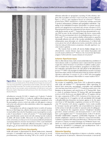
Figure 49–6. Electron micrograph of negatively stained fiber of HgS Activation of the Coagulation System
and the structure deduced by three-dimensional image reconstruction. The initiator of coagulation, tissue factor (TF), is elevated in patients
The reconstructed fiber is presented as ball models, with each ball rep- with sickle cell anemia. 40,74,85–87 Microparticles (MPs) expressing TF
resenting a HgS tetramer. The models are presented as the outer sheath derived from monocytes, macrophages, neutrophils and endothelial
(left), the inner core (center), and a combination of both inner and outer cells have been described in SCD. 58,68,74,88 Conflicting results exist in the
filaments (right). (Reproduced with permission from the University of Texas literature on the presence and contribution of TF bearing MPs. There
Medical Branch.) is a lack of correlation between TF bearing MPs and procoagulant
activity in SCD. Erythrocyte and platelet MPs are TF-negative and are
cell adhesion molecule (VCAM)-1, integrin α β , P-selectin, P-selectin the major component of MPs in SCD. Activation of the intrinsic path-
V 3
glycoprotein ligand (PSGL)-1, E-selectin, Lutheran blood group anti- way of coagulation by TF-negative, red cell, and platelet MPs through
gen, and thrombospondin. 55–60 The site of adhesion is purported to be a phosphatidylserine-dependent mechanism appears to be the major
the postcapillary venule at which site sickle red cells appear to interact contributor of MP-dependent coagulation activation in SCD. Perivas-
with white cells adherent to the endothelium rather than engaging the cular TF interaction with plasma coagulation factors made possible
endothelium directly. 31 by increased vascular permeability and phosphatidylserine exposure
Neutrophilia is an adverse prognostic factor in sickle cell anemia. on the surface of red cells secondary to repeated cycles of sickling pro-
Because of their larger size, adherent leukocytes cause a greater decrease vide an impetus for the coagulation process. Heightened thrombin
89
in vessel caliber than red cells. Diapedesis occurs in postcapillary venules, generation, platelet activation, and decreased protein C and S levels
a site of vasoocclusion in sickle cell anemia. 31,61–63 Neutrophil integrin favor a procoagulant state. 69,90,91 Increased plasma levels of D-dimers,
α β microdomains capture sickle red cells causing vascular occlusion thrombin–antithrombin complexes, prothrombin fragment 1.2, and
M 2
in sickle cell mouse models. Monocytes are also highly activated in plasmin–antiplasmin complexes are indicative of increased thrombin-
sickle cell anemia, and they promote increased endothelial activation by mediated coagulation with subsequent fibrinolysis. Plasma from sickle
92
increased production of tumor necrosis factor (TNF)-α and interleukin cell patients contains increased ultralarge von Willebrand factor mul-
(IL)-1β. Expression of leukocyte adhesion molecules, L-selectin, and timers as a result of increased endothelial cell secretion and impaired
60
integrin α β , are associated with a severe clinical phenotype. 61,64 cleavage by ADAMTS13 (a disintegrin and metalloprotease with a
M 2
thrombospondin type 1 motif member 13). 93
Inflammation and Chronic Vasculopathy
Sickle cell anemia is characterized by chronic leukocytosis, abnormal Adenosine Signaling
activation of neutrophils and monocytes, and an increase in several Cellular stress leads to the degradation of adenine nucleotides, resulting
proinflammatory mediators including TNF-α, IL-6, and IL-1β. Several in the generation of adenosine. Adenosine homeostasis is maintained
Kaushansky_chapter 49_p0759-0788.indd 765 9/18/15 3:01 PM