
Page 787 - Williams Hematology ( PDFDrive )
P. 787
762 Part VI: The Erythrocyte Chapter 49: Disorders of Hemoglobin Structure: Sickle Cell Anemia and Related Abnormalities 763
NOMENCLATURE OF ABNORMAL SICKLE CELL DISEASE
HEMOGLOBINS DEFINITION AND HISTORY
Following the molecular characterization of HbS by Ingram and col- The first case of SCD, reported in 1910, was that of a dental student from
leagues in 1956, there has been a rapid and exponential increase in Grenada, Walter Clement-Noel, studying in Chicago. Dr. James Her-
the number of variant or “abnormal” Hbs. This number now exceeds rick and his intern, Dr. Ernest Irons, were in charge of Mr. Noel’s care
1
1000. A detailed description of variant Hbs, their chemical and func- between 1904 and 1907, during which time he had several bouts of fever
tional properties, and population distribution can be found on the Glo- and cough and a history of leg ulcers, jaundice, and exercise intolerance.
bin Gene Server website (http://globin.cse.psu.edu/). Initially, newly Herrick and Irons made astute clinical observations and prepared blood
described variants were designated by letters of the alphabet (e.g., HbC, films and photomicrographs of nucleated red blood cells and of red cells
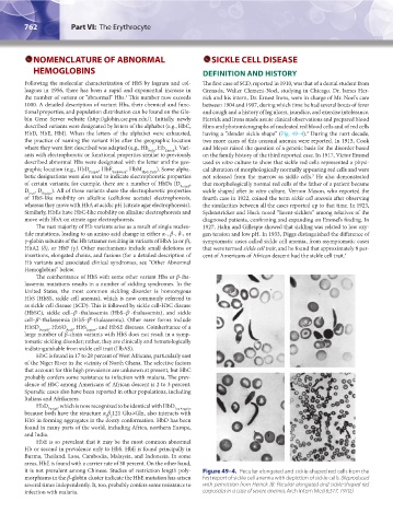
HbD, HbE, HbJ). When the letters of the alphabet were exhausted, having a “slender sickle shape” (Fig. 49–4). During the next decade,
2
the practice of naming the variant Hbs after the geographic location two more cases of this unusual anemia were reported. In 1915, Cook
where they were first described was adapted (e.g., Hb Koln , Hb Zurich ). Vari- and Meyer raised the question of a genetic basis for the disorder based
ants with electrophoretic or functional properties similar to previously on the family history of the third reported case. In 1917, Victor Emmel
described abnormal Hbs were designated with the letter and the geo- used in vitro culture to show that sickle red cells represented a physi-
graphic location (e.g., HbD Punjab , HbE Saskatoon , HbM Hyde Park ). Some alpha- cal alteration of morphologically normally appearing red cells and were
betic designations were also used to indicate electrophoretic properties not released from the marrow as sickle cells. He also demonstrated
3
of certain variants; for example, there are a number of HbDs (D Punjab , that morphologically normal red cells of the father of a patient became
D , D Ibadan ). All of these variants share the electrophoretic properties sickle shaped after in vitro culture. Vernon Mason, who reported the
Iran
of HbS-like mobility on alkaline (cellulose acetate) electrophoresis, fourth case in 1922, coined the term sickle cell anemia after observing
whereas they move with HbA at acidic pH (citrate agar electrophoresis). the similarities between all the cases reported up to that time. In 1923,
Similarly, HbEs have HbC-like mobility on alkaline electrophoresis and Sydenstricker and Huck noted “latent-sicklers” among relatives of the
move with HbA on citrate agar electrophoresis. diagnosed patients, confirming and expanding on Emmel’s finding. In
The vast majority of Hb variants arise as a result of single nucleo- 1927, Hahn and Gillespie showed that sickling was related to low oxy-
tide mutations, leading to an amino acid change in either α-, β-, δ-, or gen tension and low pH. In 1933, Diggs distinguished the difference of
γ-globin subunits of the Hb tetramer resulting in variants of HbA (α or β), symptomatic cases called sickle cell anemia, from asymptomatic cases
HbA2 (δ), or HbF (γ). Other mechanisms include small deletions or that were termed sickle cell trait, and he found that approximately 8 per-
insertions, elongated chains, and fusions (for a detailed description of cent of Americans of African descent had the sickle cell trait. 4
Hb variants and associated clinical syndromes, see “Other Abnormal
Hemoglobins” below.
The coinheritance of HbS with some other variant Hbs or β-tha-
lassemia mutations results in a number of sickling syndromes. In the
United States, the most common sickling disorder is homozygous
HbS (HbSS, sickle cell anemia), which is now commonly referred to
as sickle cell disease (SCD). This is followed by sickle cell-HbC disease
(HbSC), sickle cell–β -thalassemia (HbS–β -thalassemia), and sickle
+
+
cell–β -thalassemia (HbS–β -thalassemia). Other rarer forms include
0
0
HbSD Punjab , HbSO Arab , HbS Lepore , and HbSE diseases. Coinheritance of a
large number of β-chain variants with HbS does not result in a symp-
tomatic sickling disorder; rather, they are clinically and hematologically
indistinguishable from sickle cell trait (HbAS).
HbC is found in 17 to 28 percent of West Africans, particularly east
of the Niger River in the vicinity of North Ghana. The selective factors
that account for this high prevalence are unknown at present, but HbC
probably confers some resistance to infection with malaria. The prev-
alence of HbC among Americans of African descent is 2 to 3 percent.
Sporadic cases also have been reported in other populations, including
Italians and Afrikaners.
HbD Punjab , which is now recognized to be identical with HbD Los Angeles
because both have the structure α β 121 Glu→Gln, also interacts with
2 2
HbS in forming aggregates in the deoxy conformation. HbD has been
found in many parts of the world, including Africa, northern Europe,
and India.
HbE is so prevalent that it may be the most common abnormal
Hb or second in prevalence only to HbS. HbE is found principally in
Burma, Thailand, Laos, Cambodia, Malaysia, and Indonesia. In some
areas, HbE is found with a carrier rate of 30 percent. On the other hand,
it is not prevalent among Chinese. Studies of restriction length poly- Figure 49–4. Peculiar elongated and sickle-shaped red cells from the
morphisms in the β-globin cluster indicate the HbE mutation has arisen first report of sickle cell anemia with depiction of sickle cells. (Reproduced
several times independently. It, too, probably confers some resistance to with permission from Herrick JB: Peculiar elongated and sickle-shaped red
infection with malaria. corpuscles in a case of severe anemia. Arch Intern Med 6:517, 1910.)
Kaushansky_chapter 49_p0759-0788.indd 762 9/18/15 3:01 PM