
Page 816 - Williams Hematology ( PDFDrive )
P. 816
790 Part VI: The Erythrocyte Chapter 50: Methemoglobinemia and Other Dyshemoglobinemias 791
This histidine, residue 87 in the α chain and 92 in the β chain, is desig-
nated as the proximal histidine. On the opposite side of the porphyrin
ring the iron atom lies adjacent to another histidine residue to which,
however, it is not covalently bonded. This distal histidine occupies posi-
A Fe B
tion 58 in the α chain and position 63 in the β chain. Under normal
circumstances oxygen is occasionally discharged from the heme pocket
as a superoxide anion, removing an electron from the iron and leaving
A it in the ferric state. The enzymatic machinery of the red cell efficiently
reduces the iron to the divalent form, converting the methemoglobin to
hemoglobin (Chap. 47).
In most of the hemoglobins M, tyrosine has been substituted for
either the proximal or the distal histidine. Tyrosine can form an iron–
phenolate complex that resists reduction to the divalent state by the
normal metabolic systems of the erythrocyte. Four hemoglobins M are
A Fe O 2 B a consequence of substitution of tyrosine for histidine in the proximal
and distal sites of the α and β chains. As Table 50–2 shows, these four
hemoglobins M have been designated by the geographic names of their
discovery, Boston, Saskatoon, Iwate, and Hyde Park.
B
Analogous His→Tyr substitutions in the γ chain of fetal hemoglo-
bin have also been documented and have been designated hemoglobin
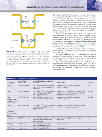
Figure 50–1. Diagrammatic representation of the heme group 57 58
inserted into the heme pocket. A, Proximal histidine; B, distal histidine. FM Osaka and FM Fort Ripley .
A. In the deoxygenated form the larger ferrous atom lies out of the Another hemoglobin M, HbM Milwaukee , is formed by substitution of
place of the porphyrin ring. B. In the oxygenated form the now smaller glutamic acid for valine in the 67th residue of the β chain. The glutamic
“ferric-like” atom can slip into the plane of the porphyrin ring. As a acid side chain points toward the heme group and its γ-carboxyl group
result, the proximal histidine, and helix F into which it is incorporated, interacts with the iron atom, stabilizing it in the ferric state.
are displaced. (Reproduced with permission from Lehmann H, It is rare for methemoglobinemia to occur as a result of hemoglo-
Huntsman RG: Man’s Haemoglobins. Philadelphia PA: Lippincott Williams & binopathies other than hemoglobins M, but Hb (β28 Leu→Met) is
Chile
Wilkins; 1974.) such a hemoglobin. Producing hemolysis only with drug administra-
tion, this unstable hemoglobin is characterized clinically by chronic
methemoglobinemia. 59
TABLE 50–2. Properties of Hemoglobins M
Amino Acid Oxygen Dissociation and Other
Hemoglobin Substitution Properties Clinical Effect Reference
HbM α58 (E7)His→Tyr Very low O affinity, almost nonex- Cyanosis resulting from formation of 182
Boston 2
istent heme–heme interaction, no methemoglobin
Bohr effect
HbM β63 (E7)His→Tyr Increased O affinity, reduced heme- Cyanosis resulting from methemoglobin for- 182,183
Saskatoon 2
heme interaction, normal Bohr mation, mild hemolytic anemia exacerbated
effect, slightly unstable by ingestion of sulfonamides
HbM α87 (F8)His→Tyr Low O affinity, negligible heme- Cyanosis resulting from formation of 182,184
Iwate 2
(HbMKankakee, heme interaction, no Bohr effect methemoglobin
HbMOldenburg,
HbMSendai)
HbM β92 (F8)His→Tyr Increased O affinity, reduced heme Cyanosis resulting from formation of methe- 79
Hyde Park 2
interaction, normal Bohr effect, moglobin, mild hemolytic anemia
slightly unstable
Hb M(hyde
park)
(HbMilwaukee 2)
HbM
Akita
HbM β67 (E11)Val →Glu Low O affinity, reduced heme-heme Cyanosis resulting from methemoglobin 185
Milwaukee 2
interaction, normal Bohr effect, formation
slightly unstable
HbFM G γ63His→Tyr Low O affinity, increased Bohr Cyanosis at birth 57
Osaka 2
effect, methemoglobinemia
HbFM G γ92His→Tyr Slightly increased O affinity Cyanosis at birth 186
Fort Ripley 2
Kaushansky_chapter 50_p0789-0800.indd 791 9/17/15 2:38 PM