
Page 965 - Williams Hematology ( PDFDrive )
P. 965
940 Part VII: Neutrophils, Eosinophils, Basophils, and Mast Cells Chapter 61: Production, Distribution, and Fate of Neutrophils 941
22,000 relative molecular mass (Mr) glycoprotein that stimulates the NEUTROPHIL KINETICS
production of neutrophils, monocytes, and eosinophils. Granulocyte Methods used to study granulocyte kinetics can be categorized as fol-
colony-stimulating factor (G-CSF) has a Mr of 20,000 and stimulates lows: (1) neutrophil depletion or destruction to determine the size and
exclusively the production of neutrophils. IL-3, or multi-CSF, also rate of mobilization of reserves and the level of compensatory neu-
has a Mr of 20,000 and acts relatively early in hematopoiesis, affect- trophil production; (2) use of radioactive tracers to study neutrophil
ing pluripotential stem cells. Finally, stem cell factor (also known as distribution, production rates, and survival times; (3) mitotic indices of
c-kit ligand or steel factor), with a Mr of 28,000, acts in combination marrow granulocytic cells to assess proliferative activity and cell cycle
with IL-3 and/or GM-CSF to stimulate the proliferation of the early times; and (4) induced inflammatory lesions to study cell movement
hematopoietic progenitor cells, basophils and mast cells. into the tissues. Of these categories, the most popular has been the use
In addition to their effects on neutrophil precursors, G-CSF and of radioactive tracers.
GM-CSF act directly on the neutrophil, enhancing its function. These Neutrophil production and neutrophil kinetics usually are ana-
cytokines regulate the production, survival, and functional activity of lyzed by describing neutrophil movement through a number of inter-
neutrophils. 21,22,26,27 In a murine model of severe bacterial infection, connected compartments. These compartments can be arranged into
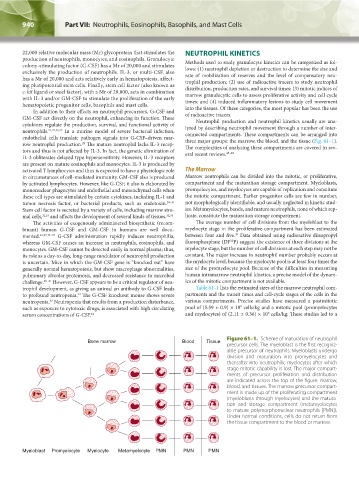
endothelial cells translate pathogen signals into G-CSF–driven mar- three major groups: the marrow, the blood, and the tissue (Fig. 61–1).
28
row neutrophil production. The mature neutrophil lacks IL-3 recep- The complexities of analyzing these compartments are covered in sev-
tors and thus is not affected by IL-3. In fact, the genetic elimination of eral recent reviews. 45–48
IL-3 obliterates delayed type hypersensitivity. However, IL-3 receptors
are present on mature eosinophils and monocytes. IL-3 is produced by
activated T lymphocytes and thus is expected to have a physiologic role The Marrow
in circumstances of cell-mediated immunity. GM-CSF also is produced Marrow neutrophils can be divided into the mitotic, or proliferative,
by activated lymphocytes. However, like G-CSF, it also is elaborated by compartment and the maturation storage compartment. Myeloblasts,
mononuclear phagocytes and endothelial and mesenchymal cells when promyelocytes, and myelocytes are capable of replication and constitute
these cell types are stimulated by certain cytokines, including IL-1 and the mitotic compartment. Earlier progenitor cells are few in number,
tumor necrosis factor, or bacterial products, such as endotoxin. 29–31 not morphologically identifiable, and usually neglected in kinetic stud-
Stem cell factor is secreted by a variety of cells, including marrow stro- ies. Metamyelocytes, bands, and mature neutrophils, none of which rep-
mal cells, 32,33 and affects the development of several kinds of tissues. 32,34 licate, constitute the maturation storage compartment.
The activities of exogenously administered biosynthetic (recom- The average number of cell divisions from the myeloblast to the
binant) human G-CSF and GM-CSF in humans are well docu- myelocyte stage in the proliferative compartment has been estimated
49
mented. 22,27,35–37 G-CSF administration rapidly induces neutrophilia, between four and five. Data obtained using radioactive diisopropyl
32
whereas GM-CSF causes an increase in neutrophils, eosinophils, and fluorophosphate (DF P) suggest the existence of three divisions at the
monocytes. GM-CSF cannot be detected easily in normal plasma; thus, myelocyte stage, but the number of cell divisions at each step may not be
its role as a day-to-day, long-range modulator of neutrophil production constant. The major increase in neutrophil number probably occurs at
is uncertain. Mice in which the GM-CSF gene is “knocked out” have the myelocyte level, because the myelocyte pool is at least four times the
generally normal hematopoiesis, but show macrophage abnormalities, size of the promyelocyte pool. Because of the difficulties in measuring
pulmonary alveolar proteinosis, and decreased resistance to microbial human intramarrow neutrophil kinetics, a precise model of the dynam-
challenge. 38–41 However, G-CSF appears to be a critical regulator of neu- ics of the mitotic compartment is not available.
trophil development, as giving an animal an antibody to G-CSF leads Table 61–1 lists the estimated sizes of the marrow neutrophil com-
to profound neutropenia. The G-CSF knockout mouse shows severe partments and the transit times and cell-cycle stages of the cells in the
42
43
neutropenia. Neutropenia that results from a production disturbance, various compartments. Precise studies have measured a postmitotic
9
such as exposure to cytotoxic drugs, is associated with high circulating pool of (5.59 ± 0.9) × 10 cells/kg and a mitotic pool (promyelocytes
9
serum concentrations of G-CSF. 44 and myelocytes) of (2.11 ± 0.36) × 10 cells/kg. These studies led to a
Figure 61–1. Scheme of maturation of neutrophil
Bone marrow Blood Tissue
precursor cells. The myeloblast is the first recogniz-
able precursor of neutrophils. Myeloblasts undergo
division and maturation into promyelocytes and
thereafter into neutrophilic myelocytes after which
stage mitotic capability is lost. The major compart-
ments of precursor proliferation and distribution
are indicated across the top of the figure: marrow,
blood, and tissues. The marrow precursor compart-
ment is made up of the proliferating compartment
(myeloblasts through myelocytes) and the matura-
tion and storage compartment (metamyelocytes
to mature polymorphonuclear neutrophils [PMN]).
Under normal conditions, cells do not return from
the tissue compartment to the blood or marrow.
Myeloblast Promyelocyte Myelocyte Metamyelocyte PMN PMN PMN
Kaushansky_chapter 61_p0939-0946.indd 940 9/18/15 9:41 AM