
Page 1059 - Clinical Immunology_ Principles and Practice ( PDFDrive )
P. 1059
1022 Part seven Organ-Specific Inflammatory Disease
anti-SMA. Also, AIH-1 may deteriorate despite corticosteroid TABLE 76.1 Diagnostic Criteria for
2
treatment and is more likely to progress to liver transplantation. autoimmune Hepatitis
Type 2 AIH (AIH-2) affects primarily girls and young women,
and it has been linked to alleles encoding the DR3 (DRB1*0301) Criteria Points
and DR7 (DRB1*07:01) molecules, and to positivity for anti-LMK Sex
antibodies. 2 Male +2
Blood tests usually show marked elevation of aminotransfer- Female 0
ases, usually with an increase in the cholestatic pattern (increased Ratio of alkaline phosphatase vs. AST/ALT
alkaline phosphatase, bilirubin). Furthermore, serum globulins, >3.0 +3
particularly of the γ type, are commonly increased in AIH cases, 1.5–2.0 +2
+1
1.0–1.5
regardless of the histological stage. <1.0 0
Lastly and most importantly, serum autoantibodies are invari- Autoantibodies (ANA, SMA, LKM-1) titer
ably positive (defined as titer >1 : 80) in patients with AIH. >1 : 80 +3
Antinuclear antibodies (ANAs) and/or antismooth-muscle (SMAs, 1 : 80 +2
directed against actin) autoantibodies are more frequently detected 1 : 40 +1
in AIH-1, whereas anti-LKM-1 antibodies (directed at cytochrome <1 : 40 0
AMA
P450 2D6 or UDP-glucuronosyltransferases) predominate in Positive −4
AIH-2. Negative 0
Without treatment, the clinical course of AIH is characterized Seropositivity for other autoantibodies +2
by high mortality, with 5- and 10-year survival rates estimated Viral hepatitis markers
as 50% and 10%, respectively. However, the use of corticosteroids Negative +3
has dramatically improved the course of the disease, with a 10-year Positive −3
2
survival rate exceeding 90%. Complications associated with AIH History of drug use −4
Yes
are similar to those of other progressive liver diseases, as chronic No +1
hepatitis can evolve to cirrhosis and ultimately to hepatocellular Average alcohol consumption (g/day)
carcinoma (HCC), despite the use of immunosuppressive therapy. <25 +2
At the time of diagnosis, approximately 30% of adult patients >60 −2
have histological evidence of cirrhosis; when appropriately treated, Presence of genetic factors (HLA, DR3 or DR4) +1
however, only a small number of patients develop cirrhosis during Presence of other autoimmune disorders +2
Liver histology
follow-up if inflammation is resolved. The occurrence of HCC Interface hepatitis +3
in patients with AIH is a rare event and develops only in long- Predominant lymphocytic infiltrate +1
standing cirrhosis. In the absence of solid data on large numbers Rosetting of liver cells +1
of cases, the incidence of primary liver neoplasia should be None of the above −5
regarded as similar to other nonviral cases of cirrhosis. Biliary changes −3
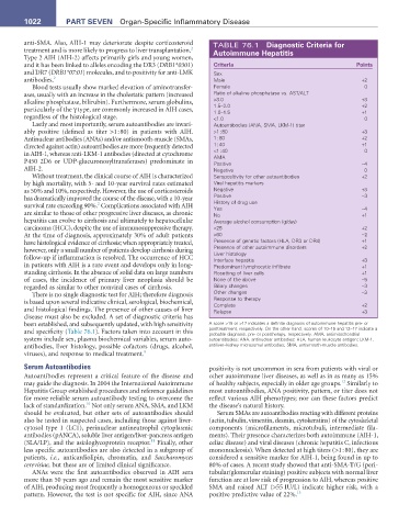
There is no single diagnostic test for AIH; therefore diagnosis Other changes −3
is based upon several indicative clinical, serological, biochemical, Response to therapy +2
Complete
and histological findings. The presence of other causes of liver Relapse +3
disease must also be excluded. A set of diagnostic criteria has
been established, and subsequently updated, with high sensitivity A score >15 or >17 indicates a definite diagnosis of autoimmune hepatitis pre- or
and specificity (Table 76.1). Factors taken into account in this posttreatment, respectively. On the other hand, scores of 10–15 and 12–17 indicate a
probable diagnosis, pre- or posttherapy, respectively. AMA, antimitochondrial
system include sex, plasma biochemical variables, serum auto- autoantibodies; ANA, antinuclear antibodies; HLA, human leukocyte antigen; LKM-1,
antibodies, liver histology, possible cofactors (drugs, alcohol, antiliver–kidney microsomal antibodies; SMA, antismooth-muscle antibodies.
viruses), and response to medical treatment. 9
Serum Autoantibodies positivity is not uncommon in sera from patients with viral or
Autoantibodies represent a critical feature of the disease and other autoimmune liver diseases, as well as in as many as 15%
12
may guide the diagnosis. In 2004 the International Autoimmune of healthy subjects, especially in older age groups. Similarly to
Hepatitis Group established procedures and reference guidelines most autoantibodies, ANA positivity, pattern, or titer does not
for more reliable serum autoantibody testing to overcome the reflect various AIH phenotypes; nor can these factors predict
10
lack of standardization. Not only serum ANA, SMA, and LKM the disease’s natural history.
should be evaluated, but other sets of autoantibodies should Serum SMAs are autoantibodies reacting with different proteins
also be tested in suspected cases, including those against liver- (actin, tubulin, vimentin, desmin, cytokeratins) of the cytoskeletal
cytosol type 1 (LC1), perinuclear antineutrophil cytoplasmic components (microfilaments, microtubuli, intermediate fila-
antibodies (pANCA), soluble liver antigen/liver-pancreas antigen ments). Their presence characterizes both autoimmune (AIH-1,
11
(SLA/LP), and the asialoglycoprotein receptor. Finally, other celiac disease) and viral diseases (chronic hepatitis C, infectious
less specific autoantibodies are also detected in a subgroup of mononucleosis). When detected at high titers (>1 : 80), they are
patients, i.e., anticardiolipin, chromatin, and Saccharomyces considered a sensitive marker for AIH-1, being found in up to
cerevisiae, but these are of limited clinical significance. 80% of cases. A recent study showed that anti-SMA-T/G (peri-
ANAs were the first autoantibodies observed in AIH sera tubular/glomerular staining) positive subjects with normal liver
more than 50 years ago and remain the most sensitive marker function are at low risk of progression to AIH, whereas positive
of AIH, producing most frequently a homogeneous or speckled SMA and raised ALT (>55 IU/L) indicate higher risk, with a
pattern. However, the test is not specific for AIH, since ANA positive predictive value of 22%. 13