
Page 1062 - Clinical Immunology_ Principles and Practice ( PDFDrive )
P. 1062
CHaPter 76 Inflammatory Hepatobiliary Diseases 1025
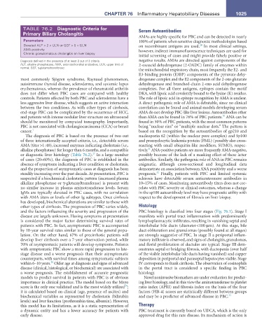
TABLE 76.2 Diagnostic Criteria for Serum Autoantibodies
Primary Biliary Cholangitis
AMAs are highly specific for PBC and can be detected in nearly
Parameters 100% of patients when sensitive diagnostic methodologies based
Elevated ALP > 2 × ULN or GGT > 5 × ULN on recombinant antigens are used. In most clinical settings,
24
AMA positivity however, indirect immunofluorescence techniques are used for
Chronic granulomatous cholangitis on liver biopsy
initial screening of cases and might provide falsely positive or
Diagnosis defined in the presence of at least 2 out of 3 criteria. negative results. AMAs are directed against components of the
ALP, alkaline phosphatase; AMA, antimitochondrial antibodies; ULN, upper limit of 2-oxoacid dehydrogenase (2-OADC) family of enzymes within
normal; GGT, γ-glutamyltransferase.
the mitochondrial respiratory chain, most frequently the E2- and
E3-binding protein (E3BP) components of the pyruvate dehy-
most commonly Sjögren syndrome, Raynaud phenomenon, drogenase complex and the E2 components of the 2-oxo glutarate
autoimmune thyroid disease, scleroderma, and systemic lupus dehydrogenase and branched-chain 2-oxo acid dehydrogenase
erythematosus, whereas the prevalence of rheumatoid arthritis complexes. For all three antigens, epitopes contain the motif
does not differ when PBC cases are compared with healthy DKA, with lipoic acid covalently bound to the lysine (K) residue.
controls. Patients affected by both PBC and scleroderma have a The role of lipoic acid in epitope recognition by AMA is unclear.
less aggressive liver disease, which suggests an active interaction A direct pathogenic role of AMA is debatable, since no clinical
between the two conditions. As with other types of cirrhosis, correlation can be found and animal models developing serum
end-stage PBC can be complicated by the occurrence of HCC, AMA do not develop PBC-like liver lesions. Autoantibodies other
30
and patients with intense nodular liver structure on ultrasound than AMA can be found in 76% of PBC patients. ANA can be
should be monitored by computed tomography. Importantly, found in 50% of PBC patients, with the most common patterns
PBC is not associated with cholangiocarcinoma (CCA) or breast being “nuclear rim” or “multiple nuclear dots.” The pattern is
cancer. 1 based on the recognition by the autoantibodies of gp210 and
The diagnosis of PBC is based on the presence of two out nucleoporin 62 (within the nuclear pore complex) and Sp100
of three internationally accepted criteria, i.e., detectable serum and promyelocytic leukemia protein (PML) (possibly also cross-
AMA (titer >1 : 40), increased enzymes indicating cholestasis (i.e., reacting with small ubiquitin-like modifiers, SUMO), respec-
31
alkaline phosphatase) for longer than 6 months, and a compatible tively. ANA-positive patients are more frequently AMA-negative,
27
or diagnostic liver histology (Table 76.2). In a large number possibly because of the lack of a masking effect of these latter
of cases (20–60%), the diagnosis of PBC is established in the antibodies. Similarly, the pathogenic role of ANA in PBC remains
absence of symptoms indicating a liver condition or cholestasis, enigmatic, although cross-sectional and longitudinal data
and the proportion of asymptomatic cases at diagnosis has been demonstrate an association between ANA positivity and a worse
32
steadily increasing over the past decade. At presentation, PBC is prognosis. Finally, patients with PBC and limited systemic
suspected if a biochemical cholestatic pattern (increased plasma sclerosis have detectable serum anticentromere antibodies in
alkaline phosphatase or γ-glutamyltransferase) is present with 10–15% of cases. Monitoring autoantibody titers does not cor-
no similar increase in plasma aminotransferase levels. Serum relate with PBC severity or clinical outcomes, whereas a change
IgMs are typically elevated in PBC cases, with no correlation in the sp100 autoantibody level may have prognostic utility with
with AMA titers or levels of other Ig subtypes. Once cirrhosis respect to the development of fibrosis on liver biopsy.
has developed, biochemical alterations are similar to those with
other types of cirrhosis. The progression of PBC varies widely, Histology
and the factors influencing the severity and progression of the PBC histology is classified into four stages (Fig. 76.1). Stage I
disease are largely unknown. Having symptoms at presentation manifests with portal tract inflammation with predominantly
is considered the major factor determining survival rates of lymphoplasmacytic infiltrates, resulting in vanishing septal and
patients with PBC. In fact, asymptomatic PBC is accompanied interlobular bile ducts (diameter <100 µm). At this stage, bile
by 10-year survival rates similar to those of the general popu- duct obliteration and granulomas (possibly found at all stages)
lation. On the other hand, 67% of precirrhotic patients will are strongly suggestive of PBC. In stage II a periportal inflam-
develop liver cirrhosis over a 7-year observation period, while matory infiltrate is observed, and signs of cholangitis, granulomas,
70% of asymptomatic patients will develop symptoms. Patients and florid proliferation of ductules are typical. Stage III dem-
with symptomatic PBC show a more rapid progression to late- onstrates septal or bridging fibrosis, with ductopenia (over half
stage disease and a worse prognosis than their asymptomatic of the visible interlobular bile ducts having vanished) and copper
counterparts, with survival times among symptomatic subjects deposition in periportal and paraseptal hepatocytes visible. Stage
28
within 6–10 years. Older age at diagnosis and signs of advanced IV corresponds to frank cirrhosis. The observation of eosinophils
disease (clinical, histological, or biochemical) are associated with in the portal tract is considered a specific finding in PBC
a worse prognosis. The establishment of accurate prognostic histology.
models to predict survival in patients with PBC is of obvious Novel noninvasive biomarkers are under evaluation for predict-
importance in clinical practice. The model based on the Mayo ing liver histology, and in this view the aminotransferase to platelet
29
score is the only one validated and is the most widely utilized ; ratio index (APRI) and fibrosis index on the basis of the four
it is calculated based on clinical (age, presence of ascites) and factors (FIB-4) scores are statistically different between groups
biochemical variables as represented by cholestasis (bilirubin and may be a predictor of advanced disease in PBC. 33
levels) and liver function (prothrombin time, albumin). However,
this model has its limitations, as it is a static representation of Therapy
a dynamic entity and has a lower accuracy for patients with PBC treatment is currently based on UDCA, which is the only
early disease. approved drug for this rare disease. Its mechanism of action is