
Page 1064 - Clinical Immunology_ Principles and Practice ( PDFDrive )
P. 1064
CHaPter 76 Inflammatory Hepatobiliary Diseases 1027
Minnesota, reported a disease prevalence of 20.9 per 100 000 normal until late stages. Autoantibodies are of limited use in
36
men and 6.3 per 100 000 women. Epidemiological data indicate the diagnosis of PSC due to low sensitivity and specificity. Only
that annual incidence rates are not increasing over time despite a limited percentage of patients (33%) has positive pANCA,
earlier ages at diagnosis, and survival is possibly longer, similar which usually is found in IBD patients without PSC.
to what was observed in PBC. Imaging represents a useful diagnostic tool, as it may find
In contrast to the vast majority of autoimmune diseases, PSC the unique strictured and dilated tracts within the intrahepatic
is more commonly diagnosed in men, with a female:male ratio or extrahepatic bile ducts. Among the imaging techniques,
estimated as 1 : 2, with a preference for 30- to 40-year-olds. PSC endoscopic retrograde cholangiopancreatography (ERCP) and
has a strong association with inflammatory bowel disease. magnetic resonance cholangiopancreatography (MRCP) are
Approximately 60–80% of patients with PSC present with currently considered equal for sensitivity, but their results are
inflammatory bowel disease, of which 87% have ulcerative colitis influenced by the operator’s skill and experience.
and 13% have Crohn disease. 37
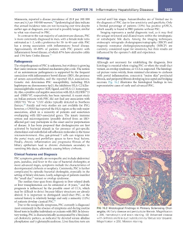
Histology
Pathogenesis Although not necessary for establishing the diagnosis, liver
The etiopathogenesis of PSC is unknown, but evidence is growing histology is essential when staging PSC or when the small-duct
that (auto) immune-mediated mechanisms play a role. The notion variant, an overlap syndrome, or CCA is suspected. The histologi-
of an autoimmune pathogenesis is supported by the frequent cal picture varies widely, from minimal alterations to cirrhosis
association with inflammatory bowel disease (IBD), the presence with portal inflammation, concentric “onion skin” periductal
of serum autoantibodies, and the reported HLA associations. fibrosis, and periportal fibrosis developing into septal and bridging
Genetic risk determines PSC susceptibility, including HLA necrosis. Fig. 76.2 illustrates the histological findings in two
haplotypes on chromosome 6p21, the inhibitory HLA-C2 killer- representative cases of early and advanced PSC.
immunoglobulin receptor (KIR) ligand, and HLA-C1 homozygos-
ity. Also, a positive and negative association with HLA-HLDRB1*15
and -DRB1*07, respectively, has been reported. A recent study
on Italian patients with PBC did not find any association with
DRB1*03, *04 or *13:01 alleles typically detected in Northern
38
Europe. Family and twin studies are not available for PSC;
however, a GWAS has reported the HLA region as the only major
association, albeit in a minority of patients and sometimes
overlapping with IBD-associated genes. The innate immune
system and microorganisms (possibly derived from an IBD-
affected gut) may participate in the onset and/or perpetuation
of disease. It has been proposed that cholangiocytes are first
activated by bacterial stimuli in the presence of gut-specific
chemokines and endothelial cell adhesion molecules in the tissue
microenvironment. Also, gut-primed T cells can migrate into
the portal tracts and peribiliary spaces to form focal lesions.
Finally, chronic inflammation and progressive fibrosis of the
biliary epithelium lead to chronic cholestasis secondary to A
vanishing bile ducts, ultimately causing biliary cirrhosis.
Clinical Features and Diagnosis
PSC symptoms generally are nonspecific and include abdominal
pain, jaundice, and fever in the case of bacterial cholangitis; at
more advanced stages, symptoms include the characteristics of
decompensated cirrhosis or neoplasia. Commonly, PSC is further
complicated by episodic bacterial cholangitis, especially in the
setting of biliary strictures. Lastly, subgroups of patients manifest
the “small duct” variant or overlap syndrome.
The median time span from diagnosis to liver-related death
39
or liver transplantation can be estimated at 18 years, and the
prognosis is influenced by the possible onset of CCA, which
may be difficult to detect because biliary structures are already
altered. It is important to distinguish small-duct PSC, as the
natural history is relatively benign and only a minority (12%)
of patients develop classical PSC. 40 B
Due to the nonspecific symptoms, PSC currently is diagnosed
most commonly in the absence of symptoms and during routine FIG 76.2 Histological Findings in Primary Sclerosing Chol-
blood tests in healthy individuals or patients with IBD. In labora- angitis. (A) Early disease and periductular fibrosis. Magnification
tory testing, PSC is characteristically accompanied by a biochemi- x 200, hematoxylin and eosin staining. (B) Advanced disease
cal cholestatic pattern, as indicated by elevated serum alkaline with cirrhosis and bile duct substitution by fibrous scar (square).
phosphatase and γ-glutamyltransferase. Liver function tests are Magnification × 200, Masson staining.