
Page 1136 - Clinical Immunology_ Principles and Practice ( PDFDrive )
P. 1136
1102 Part NiNe Transplantation
1. Normal
2. Antibody-mediated rejection
Acute antibody-mediated rejection
I. ATN-like – C4d+, minimal inflammation
II. Capillary - margination and/or thromboses, C4d+
III. Arterial – v3, C4d+
Chronic active antibody-mediated rejection
Glomerular double contours and/or peritubular capillary basement membrane multilayering
and/or interstitial fibrosis/tubular atrophy and/or fibrous intimal thickening in arteries, C4d
3. Borderline changes: ‘suspicious’ for acute T-cell-mediated rejection
This category is used when no intimal arteritis is present, but there are foci of tubulitis
4. T-cell-mediated rejection
Acute T-cell-mediated rejection
IA. Cases with significant interstitial infiltration (>25% of parenchyma affected, i2 or i3)
and foci of moderate tubulitis (t2)
IB. Cases with significant interstitial infiltration (>25% of parenchyma affected, i2 or i3)
and foci of severe tubulitis (t3)
IIA. Cases with mild to moderate intimal arteritis (v1)
IIB. Cases with severe intimal arteritis comprising >25% of the luminal area (v2)
III. Cases with ‘transmural’ arteritis and/or arterial fibrinoid change and necrosis of medial
smooth muscle cells with accompanying lymphocytic inflammation (v3)
Chronic active T-cell-mediated rejection
‘Chronic allograft arteriopathy’ (arterial intimal fibrosis with mononuclear cell infiltration in
fibrosis, formation of neo-intima)
5. Interstitial fibrosis and tubular atrophy, no evidence of any specific etiology
I. Mild interstitial fibrosis and tubular atrophy (<25% of cortical area)
II. Moderate interstitial fibrosis and tubular atrophy (26–50% of cortical area)
III. Severe interstitial fibrosis and tubular atrophy/loss (>50% of cortical area)
(may include nonspecific vascular and glomerular sclerosis, but severity
graded by tubulointerstitial features)
6. Other: Changes not considered to be due to rejection-acute and/or chronic
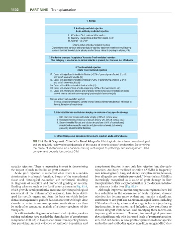
FiG 81.4 Banff Diagnostic Criteria for Renal Allografts. Histological criteria have been developed
and are regularly reviewed to aid diagnosis of the cause of chronic allograft dysfunction. Determining
the cause of dysfunction aids decision making with regard to pathology and management. C4d,
complement degradation product C4d.
vascular rejection. There is increasing interest in determining complement fixation in not only late rejection but also early
the impact of such antibodies on graft outcome. rejection. Antibody-mediated rejection (ABMR) is frequently
Acute graft rejection is suspected when there is a sudden seen following heart, lung, and kidney transplantations; however,
20
deterioration in allograft function. Biopsy of the transplanted liver allografts are relatively protected. Nevertheless ABMR is
tissue and histological evaluation are performed, resulting increasingly recognized as a cause of graft damage in liver
in diagnosis of rejection and numerical grading of severity. transplantation. This is explored further in the discussion below
Grading schemes, such as the Banff criteria shown in Fig. 81.4, on tolerance in the liver (Fig. 81.6).
which provide semiquantitative measures for histopathological Although improved immunosuppression regimens have led
assessment of the inflammatory response, have been devel- to a reduction in the occurrence of acute rejection, chronic
oped for specific organs and form the basis on which further rejection has become more evident and remains a significant
clinical management is guided; decisions to treat with high-dose contributor to late graft loss. Nonimmunological factors, including
steroids or other immunosuppressive medications can thus CNI-induced toxicity, advanced donor age, ischemic injury during
be made after structured analysis of a biopsy specimen of the implantation, hypertension, and infection, also contribute to
transplant. chronic allograft dysfunction, and modifying these factors can
21
In addition to the diagnosis of cell-mediated rejection, modern improve graft outcome. However, immunological processes
staining techniques have enabled the identification of complement play a significant role with increased levels of pretransplantation
component 4d (C4d) in biopsy specimens from rejecting tissues, anti-HLA antibodies, de novo posttransplantation donor-specific
thus providing indirect evidence of antibody deposition and antibodies and antibodies against non-HLA antigen MHC class