
Page 141 - Clinical Immunology_ Principles and Practice ( PDFDrive )
P. 141
CHaPTEr 8 T-Cell Development 123
Normal MHC Defective MHC I Normal MHC I Male-specific TCR transgene; MHC I restricted
a
expression Normal MHC II Defective MHC II
FEMALE FEMALE MALE
MHC a MHC b MHC a
DP DP DP
DP DP DP
CD4 CD8 CD4 CD8 CD4 CD8
SP SP SP SP SP SP CD4 CD8 CD4 CD8 CD4 CD8
SP SP SP SP SP SP
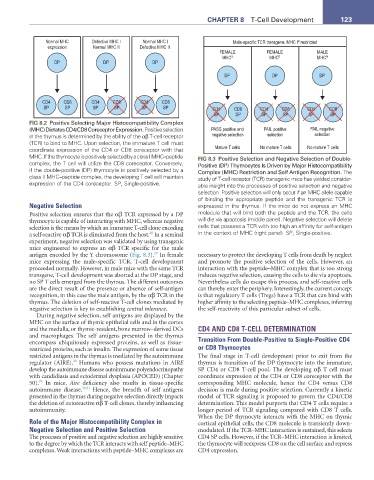
FIG 8.2 Positive Selecting Major Histocompatibility Complex
(MHC) Dictates CD4/CD8 Coreceptor Expression. Positive selection PASS positive and FAIL positive FAIL negative
in the thymus is determined by the ability of the αβ T-cell receptor negative selection selection selection
(TCR) to bind to MHC. Upon selection, the immature T cell must
coordinate expression of the CD4 or CD8 coreceptor with that Mature T cells No mature T cells No mature T cells
MHC. If the thymocyte is positively selected by a class I MHC–peptide FIG 8.3 Positive Selection and Negative Selection of Double-
complex, the T cell will utilize the CD8 coreceptor. Conversely, Positive (DP) Thymocytes Is Driven by Major Histocompatibility
if the double-positive (DP) thymocyte is positively selected by a Complex (MHC) Restriction and Self Antigen Recognition. The
class II MHC–peptide complex, the developing T cell will maintain study of T-cell receptor (TCR) transgenic mice has yielded consider-
expression of the CD4 coreceptor. SP, Single-positive. able insight into the processes of positive selection and negative
selection. Positive selection will only occur if an MHC allele capable
of binding the appropriate peptide and the transgenic TCR is
Negative Selection expressed in the thymus. If the mice do not express an MHC
Positive selection ensures that the αβ TCR expressed by a DP molecule that will bind both the peptide and the TCR, the cells
thymocyte is capable of interacting with MHC, whereas negative will die via apoptosis (middle panel). Negative selection will delete
selection is the means by which an immature T-cell clone encoding cells that possess a TCR with too high an affinity for self-antigen
20
a self-reactive αβ TCR is eliminated from the host. In a seminal in the context of MHC (right panel). SP, Single-positive.
experiment, negative selection was validated by using transgenic
mice engineered to express an αβ TCR specific for the male
27
antigen encoded by the Y chromosome (Fig. 8.3). In female necessary to protect the developing T cells from death by neglect
mice expressing the male-specific TCR, T-cell development and promote the positive selection of the cells. However, an
proceeded normally. However, in male mice with the same TCR interaction with the peptide–MHC complex that is too strong
transgene, T-cell development was aborted at the DP stage, and induces negative selection, causing the cells to die via apoptosis.
no SP T cells emerged from the thymus. The different outcomes Nevertheless cells do escape this process, and self-reactive cells
are the direct result of the presence or absence of self-antigen can thereby enter the periphery. Interestingly, the current concept
recognition, in this case the male antigen, by the αβ TCR in the is that regulatory T cells (Tregs) have a TCR that can bind with
thymus. The deletion of self-reactive T-cell clones mediated by higher affinity to the selecting peptide–MHC complexes, inferring
negative selection is key to establishing central tolerance. the self-reactivity of this particular subset of cells.
During negative selection, self antigens are displayed by the
MHC on the surface of thymic epithelial cells and in the cortex
and the medulla, or thymic-resident, bone marrow–derived DCs CD4 AND CD8 T-CELL DETERMINATION
and macrophages. The self antigens presented in the thymus
encompass ubiquitously expressed proteins, as well as tissue- Transition From Double-Positive to Single-Positive CD4
restricted proteins, such as insulin. The expression of some tissue or CD8 Thymocytes
restricted antigens in the thymus is mediated by the autoimmune The final stage in T-cell development prior to exit from the
28
regulator (AIRE). Humans who possess mutations in AIRE thymus is transition of the DP thymocyte into the immature,
develop the autoimmune disease autoimmune polyendocrinopathy SP CD4 or CD8 T-cell pool. The developing αβ T cell must
with candidiasis and ectodermal dysplasia (APOCED) (Chapter coordinate expression of the CD4 or CD8 coreceptor with the
29
50). In mice, Aire deficiency also results in tissue-specific corresponding MHC molecule, hence the CD4 versus CD8
autoimmune disease. 30,31 Hence, the breadth of self antigens decision is made during positive selection. Currently a kinetic
presented in the thymus during negative selection directly impacts model of TCR signaling is proposed to govern the CD4/CD8
the deletion of autoreactive αβ T-cell clones, thereby influencing determination. This model purports that CD4 T cells require a
autoimmunity. longer period of TCR signaling compared with CD8 T cells.
When the DP thymocyte interacts with the MHC on thymic
Role of the Major Histocompatibility Complex in cortical epithelial cells, the CD8 molecule is transiently down-
Negative Selection and Positive Selection modulated. If the TCR–MHC interaction is sustained, this selects
The processes of positive and negative selection are highly sensitive CD4 SP cells. However, if the TCR–MHC interaction is limited,
to the degree by which the TCR interacts with self peptide–MHC the thymocyte will reexpress CD8 on the cell surface and repress
complexes. Weak interactions with peptide–MHC complexes are CD4 expression.