
Page 180 - Clinical Immunology_ Principles and Practice ( PDFDrive )
P. 180
162 ParT ONE Principles of Immune Response
Endothelial cell
G
A
G
CK γ GTP
α i α 1
GDP β
Leukocyte
RGS
GRK P13 Kγ γ PLC β2/β3
Arrestin
β
PIP2 PKC
IP3 R
PIP3 DAG Ca 2+
Ca 2+
RhoGEF RacGEF cdc42GEF IP3 Ca 2+
Rho Rac cdc42
PKCζ ROCK Pak Arp2/3
myosin F-actin
Adhesion Contraction F-Actin polymerization
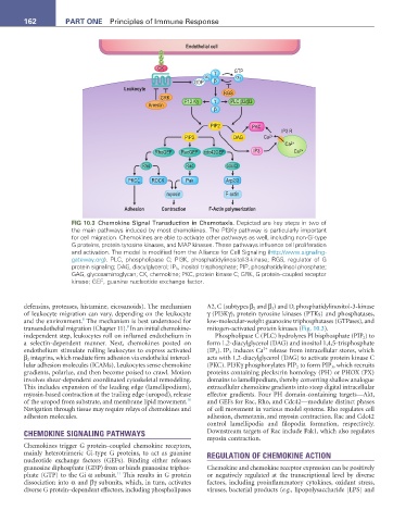
FIG 10.3 Chemokine Signal Transduction in Chemotaxis. Depicted are key steps in two of
the main pathways induced by most chemokines. The PI3Kγ pathway is particularly important
for cell migration. Chemokines are able to activate other pathways as well, including non-Gi-type
G proteins, protein tyrosine kinases, and MAP kinases. These pathways influence cell proliferation
and activation. The model is modified from the Alliance for Cell Signaling (http://www.signaling-
gateway.org). PLC, phospholipase C; PI3K, phosphatidylinositol-3-kinase; RGS, regulator of G
protein signaling; DAG, diacylglycerol; IP 3 , inositol trisphosphate; PIP, phosphatidylinsol phosphate;
GAG, glycosaminoglycan; CK, chemokine; PKC, protein kinase C; GRK, G protein–coupled receptor
kinase; GEF, guanine nucleotide exchange factor.
defensins, proteases, histamine, eicosanoids). The mechanism A2, C (subtypes β 2 and β 3 ) and D, phosphatidylinositol-3-kinase
of leukocyte migration can vary, depending on the leukocyte γ (PI3Kγ), protein tyrosine kinases (PTKs) and phosphatases,
8
and the environment. The mechanism is best understood for low-molecular-weight guanosine triphosphatases (GTPases), and
9
transendothelial migration (Chapter 11). In an initial chemokine- mitogen-activated protein kinases (Fig. 10.3).
independent step, leukocytes roll on inflamed endothelium in Phospholipase C (PLC) hydrolyzes PI bisphosphate (PIP 2 ) to
a selectin-dependent manner. Next, chemokines posted on form 1,2-diacylglycerol (DAG) and inositol 1,4,5-trisphosphate
2+
endothelium stimulate rolling leukocytes to express activated (IP 3). IP 3 induces Ca release from intracellular stores, which
β 2 integrins, which mediate firm adhesion via endothelial intercel- acts with 1,2-diacylglycerol (DAG) to activate protein kinase C
lular adhesion molecules (ICAMs). Leukocytes sense chemokine (PKC). PI3Kγ phosphorylates PIP 2 to form PIP 3 , which recruits
gradients, polarize, and then become poised to crawl. Motion proteins containing pleckstrin homology (PH) or PHOX (PX)
involves shear-dependent coordinated cytoskeletal remodeling. domains to lamellipodium, thereby converting shallow analogue
This includes expansion of the leading edge (lamellipodium), extracellular chemokine gradients into steep digital intracellular
myosin-based contraction at the trailing edge (uropod), release effector gradients. Four PH domain-containing targets—Akt,
10
of the uropod from substrate, and membrane lipid movement. and GEFs for Rac, Rho, and Cdc42—modulate distinct phases
Navigation through tissue may require relays of chemokines and of cell movement in various model systems. Rho regulates cell
adhesion molecules. adhesion, chemotaxis, and myosin contraction. Rac and Cdc42
control lamellipodia and filopodia formation, respectively.
CHEMOKINE SIGNALING PATHWAYS Downstream targets of Rac include Pak1, which also regulates
myosin contraction.
Chemokines trigger G protein–coupled chemokine receptors,
mainly heterotrimeric Gi-type G proteins, to act as guanine REGULATION OF CHEMOKINE ACTION
nucleotide exchange factors (GEFs). Binding either releases
guanosine diphosphate (GDP) from or binds guanosine triphos- Chemokine and chemokine receptor expression can be positively
11
phate (GTP) to the Gi α subunit. This results in G protein or negatively regulated at the transcriptional level by diverse
dissociation into α and βγ subunits, which, in turn, activates factors, including proinflammatory cytokines, oxidant stress,
diverse G protein–dependent effectors, including phospholipases viruses, bacterial products (e.g., lipopolysaccharide [LPS] and