
Page 191 - Clinical Immunology_ Principles and Practice ( PDFDrive )
P. 191
172 ParT ONE Principles of Immune Response
Skin
Thoracic duct
Afferent lymphatic Postcapillary
vessels venule
Lymph node Heart
Arteries
Efferent
lymphatic PP
vessel
Gut A
FIG 11.1 Lymphocyte Recirculation Routes Under Physiologi
cal Conditions. A low level of continuous antigenic transport
into lymphoid organs takes place via the afferent lymphatics
draining the skin and epithelium of the gut. Bloodborne lympho
cytes enter the organized lymphatic tissues (lymph nodes and
Peyer patches [PP]) from the circulation via the arterial tree,
flow through the capillary bed, and then extravasate in the
postcapillary high endothelial venules (HEVs). The extravasated
lymphocytes percolate through the tissue parenchyma, enter
the lymphatic vessels, and are then carried via the efferent
lymphatics back to the systemic circulation. (Most of the venous
circulation has been omitted from the figure.) Inset: an HEV.
(From Salmi M, Jalkanen S. How do lymphocytes know where
to go: current concepts and enigmas of lymphocyte homing.
Adv Immunol 1997;64:139, with permission from Elsevier.) B
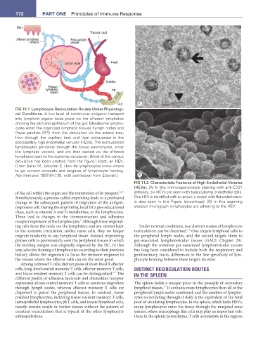
FIG 11.2 Characteristic Features of High Endothelial Venules
(HEVs). (A) In this immunoperoxidase staining with antiCD31
of the cell within the organ and the maturation of its progeny. 2,6,7 antibody, six HEVs are seen with typical plump endothelial cells.
Simultaneously, a process called imprinting leads to a profound One HEV is identified with an arrow; a vessel with flat endothelium
change in the subsequent pattern of migration of the antigen- is also seen in this Figure (arrowhead). (B) In this scanning
responsive cell. During the imprinting, local DCs give educational electron micrograph lymphocytes are adhering to the HEV.
clues, such as vitamin A and D metabolites, to the lymphocytes.
These lead to changes in the chemoattractant and adhesion
8
receptor repertoire of the lymphocytes. Although these respond-
ing cells leave the node via the lymphatics and are carried back Under normal conditions, two distinct routes of lymphocyte
2,6
to the systemic circulation, unlike naïve cells, they no longer recirculation can be discerned. One targets lymphoid cells to
migrate randomly to any lymphoid tissue. Instead, imprinting the peripheral lymph nodes, and the second targets them to
primes cells to preferentially seek the peripheral tissues in which gut-associated lymphoreticular tissues (GALT; Chapter 20).
the inciting antigen was originally ingested by the DC. In this Although the common gut-associated lymphoreticular system
way, selective homing of lymphocytes according to their previous has long been considered to include both the respiratory and
history allows the organism to focus the immune response to genitourinary tracts, differences in the fine specificity of lym-
the tissues where the effector cells can do the most good. phocyte homing between these targets do exist.
Among activated T cells, distinct pools of short-lived T effector
cells, long-lived central memory T cells, effector memory T cells, DISTINCT RECIRCULATION ROUTES
6,9
and tissue resident memory T cells can be distinguished. The IN THE SPLEEN
different profile of adhesion molecule and chemokine receptor
expression allows central memory T cells to continue migration The spleen holds a unique place in the panoply of secondary
10
through lymph nodes, whereas effector memory T cells are lymphoid tissues. It contains more lymphocytes than all of the
dispersed to patrol the peripheral tissues. In contrast, tissue peripheral lymph nodes combined, and the number of lympho-
resident lymphocytes, including tissue resident memory T cells, cytes recirculating through it daily is the equivalent of the total
intraepithelial lymphocytes, γδ T cells, and innate lymphoid cells, pool of circulating lymphocytes. In the spleen, which lacks HEVs,
mostly remain sessile in barrier tissues without the pattern of many lymphocytes enter the tissue through the marginal zone
constant recirculation that is typical of the other lymphocyte sinuses, where macrophage-like cells may play an important role.
subpopulations. Once in the splenic parenchyma, T cells accumulate in the regions