
Page 193 - Clinical Immunology_ Principles and Practice ( PDFDrive )
P. 193
174 ParT ONE Principles of Immune Response
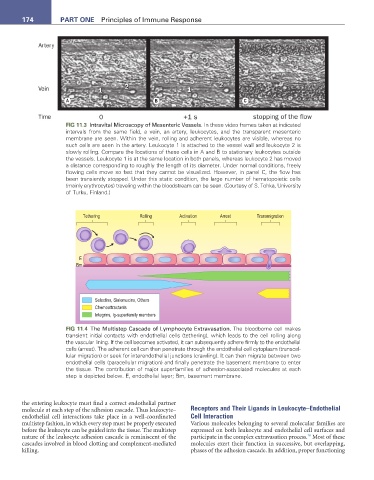
A B C
FIG 11.3 Intravital Microscopy of Mesenteric Vessels. In these video frames taken at indicated
intervals from the same field, a vein, an artery, leukocytes, and the transparent mesenteric
membrane are seen. Within the vein, rolling and adherent leukocytes are visible, whereas no
such cells are seen in the artery. Leukocyte 1 is attached to the vessel wall and leukocyte 2 is
slowly rolling. Compare the locations of these cells in A and B to stationary leukocytes outside
the vessels. Leukocyte 1 is at the same location in both panels, whereas leukocyte 2 has moved
a distance corresponding to roughly the length of its diameter. Under normal conditions, freely
flowing cells move so fast that they cannot be visualized. However, in panel C, the flow has
been transiently stopped. Under this static condition, the large number of hematopoietic cells
(mainly erythrocytes) traveling within the bloodstream can be seen. (Courtesy of S. Tohka, University
of Turku, Finland.)
Tethering Rolling Activation Arrest Transmigration
E
Bm
Selectins, Sialomucins, Others
Chemoattractants
Integrins, Ig-superfamily members
FIG 11.4 The Multistep Cascade of Lymphocyte Extravasation. The bloodborne cell makes
transient initial contacts with endothelial cells (tethering), which leads to the cell rolling along
the vascular lining. If the cell becomes activated, it can subsequently adhere firmly to the endothelial
cells (arrest). The adherent cell can then penetrate through the endothelial cell cytoplasm (transcel
lular migration) or seek for interendothelial junctions (crawling). It can then migrate between two
endothelial cells (paracellular migration) and finally penetrate the basement membrane to enter
the tissue. The contribution of major superfamilies of adhesionassociated molecules at each
step is depicted below. E, endothelial layer; Bm, basement membrane.
the entering leukocyte must find a correct endothelial partner
molecule at each step of the adhesion cascade. Thus leukocyte– Receptors and Their Ligands in Leukocyte–Endothelial
endothelial cell interactions take place in a well-coordinated Cell Interaction
multistep fashion, in which every step must be properly executed Various molecules belonging to several molecular families are
before the leukocyte can be guided into the tissue. The multistep expressed on both leukocyte and endothelial cell surfaces and
16
nature of the leukocyte adhesion cascade is reminiscent of the participate in the complex extravasation process. Most of these
cascades involved in blood clotting and complement-mediated molecules exert their function in successive, but overlapping,
killing. phases of the adhesion cascade. In addition, proper functioning