
Page 293 - Clinical Immunology_ Principles and Practice ( PDFDrive )
P. 293
274 ParT TwO Host Defense Mechanisms and Inflammation
The dermis lies beneath the epidermis and serves as a con-
Maintenance of barrier integrity in normal skin
nective tissue layer that provides elasticity and tensile strength
to skin. It is filled with a matrix of collagen bundles, elastic fibers,
Epidermis
Commensal microorganisms AMPs glycoproteins, proteoglycans, and glycosaminoglycans, all of
Stratum corneum which are produced by dermal fibroblasts. The production of
Granular layer collagen is a dynamic process that involves continual remodeling.
Spinous layer
Langerhans Fibroblasts must balance ongoing synthesis of matrix components
cell (LC) with the production of matrix metalloproteinases (MMPs) that
Basal layer degrade the matrix. Scleroderma and morphea (localized
Melanocyte scleroderma) (Chapter 55) are inflammatory disorders that
BASEMENT MEMBRANE ZONE lead to the overproduction of collagen in the dermis, thus
Barrier integrity factors
DERMIS hardening skin.
Fibroblast Other cell types normally found in the dermis are mast cells
3
and diverse sets of tissue macrophages and dendritic cells (DCs).
Dermal DCs Langerin + The dermis supports the microvasculature of skin. Endothelial
(dDCs) dDC cells of dermal arterioles, capillaries, and postcapillary venules
pDC behave differently from those in larger vessels. They express
T cells E- and P-selectins, which enable circulating skin-homing T cells
Treg
TGF β to bind through their homing receptors cutaneous lymphocyte-
Plasmacytoid DC Langerin – TGF β IL-10 associated antigen (CLA) and P-selectin glycoprotein ligand
IL-10
pDC dDC (PSGL), respectively. 4
Trm Both homeostatic T-cell trafficking and recruitment of inflam-
Macrophages matory leukocytes into skin from the circulation require dermal
Effector endothelial cell-specific expression of E- and P-selectins. The
memory
T cells egress of leukocytes from blood occurs primarily through
postcapillary venules. Egress is regulated, in part, by endothelial
responses to cutaneous cytokines and chemokines.
Mast cells
New vessels can also develop in skin. The new vessels can
Mast cell worsen immunologically mediated dermatological diseases and
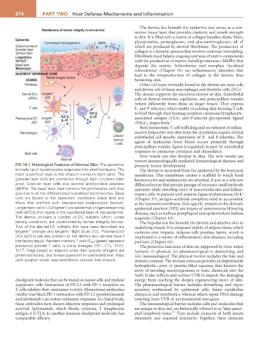
FIG 19.1 Histological Features of Normal Skin. The epidermis promote tumor development.
is made up of keratinocytes organized into stratified layers. The The dermis is separated from the epidermis by the basement
most superficial layer is the stratum corneum (light pink). The membrane. This membrane creates a scaffold to which basal
granular layer cells are connected through tight junctions (dark keratinocytes and melanocytes are attached. It acts as a selective
grey). Granular layer cells also secrete antimicrobial peptides diffusion barrier that permits passage of necessary small molecule
(AMPs). The basal layer (tan) contains the proliferative cells that nutrients while retarding entry of macromolecules and inflam-
give rise to all the differentiated suprabasal keratinocytes. Basal matory cells. In patients with systemic lupus erythematosus (SLE)
cells are bound to the basement membrane (black line) and (Chapter 51), antigen–antibody complexes tend to accumulate
share that scaffold with interspersed melanocytes (brown). at the basement membrane. Skin-specific proteins in the dermal–
Langerhans cells (LCs) (green) are epidermal antigen-presenting epidermal junction (DEJ) are targets of autoimmune blistering
cells (APCs) that reside in the suprabasal layer of the epidermis. diseases, such as bullous pemphigoid and epidermolysis bullosa
The dermis contains a number of DC subsets which, under acquisita (Chapter 63).
resting conditions, are conditioned by barrier integrity factors. The hypodermis lies beneath the dermis and attaches skin to
Two of the dermal DC subsets that have been described are underlying muscle. It is composed mainly of adipose tissue, which
−
+
langerin (orange) and langerin (light blue) DCs. Plasmacytoid cushions skin impacts. Adipose cells produce leptin, which is
DCs (pDCs) are also present in the dermis and secrete type I implicated in a variety of inflammatory skin diseases, including
interferons (aqua). Resident memory T cells (T RM ) (green) represent psoriasis (Chapter 64).
previously primed T cells in many lineages (Th1, CTL, Th17, The protective functions of skin are supported by three major
Tc17, Treg) poised to respond when their specific antigens are barriers: (i) physical, (ii) pharmacological or detoxifying, and
presented locally, but remain quiescent in uninfected skin. Mast (iii) immunological. The physical barrier includes the hair and
cells (purple) reside near endothelial venules (not shown). stratum corneum. The stratum corneum provides an impermeable
hydrophobic cover of protein-filled squames that hinders the
entry of invading microorganisms or toxic chemicals into the
body. It also reflects and scatters UVR to impede the damaging
checkpoint molecule that can be found on tumor cells and myeloid energy from reaching the deeper, regenerating layers of skin.
suppressor cells. Interaction of PD-L1 with PD-1 receptors on The pharmacological barrier includes detoxifying and repair
T cells inhibits their antitumor activity. Monoclonal antibodies enzymes synthesized by epidermal cells. Some metabolize
(mAbs) that block PD-1 interaction with PD-L1 (pembrolizumab chemicals and xenobiotics, whereas others repair DNA damage
and nivolumab) can restore antitumor responses. In clinical trials, resulting from UVR or environmental mutagens.
these antibodies have shown objective responses and prolonged The immunological barrier includes cells and molecules that
survival. Ipilimumab, which blocks cytotoxic T lymphocyte are unique to skin and are historically referred to as “skin associ-
5
antigen-4 (CTLA-4), another immune checkpoint molecule, has ated lymphoid tissue.” They include elements of both innate
comparable efficacy. immunity and acquired immunity. Together, these elements