
Page 294 - Clinical Immunology_ Principles and Practice ( PDFDrive )
P. 294
CHaPTEr 19 Host Defenses in Skin 275
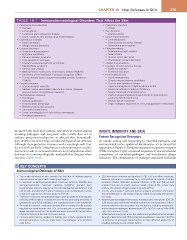
TABLE 19.1 Immunodermatological Disorders That affect the Skin
• Papulosquamous disorders • Pigmentary disorders
• Psoriasis • Vitiligo
• Lichen planus • Hair disorders
• Cutaneous graft-versus-host disease • Alopecia areata
• Acute, subacute, and discoid lupus erythematosus • Autoimmune disorders
• Eczematous disorders • Dermatomyositis
• Atopic dermatitis • Mixed connective tissue disease
• Allergic contact dermatitis • Scleroderma and morphea
• Urticarial disorders • Photodermatoses
• Urticaria and angioedema • Polymorphous light eruption
• Erythema multiforme • Solar urticaria
• Stevens Johnson syndrome • Chronic actinic dermatitis
• Toxic epidermal necrolysis • Photoallergic contact dermatitis
• Cryopyrin-associated periodic syndromes • Allergic drug eruptions
• Muckle-Wells syndrome • Disorders of subcutaneous tissues
• Familial cold urticaria • Erythema nodosum
• Neonatal-onset multisystem inflammatory disease (NOMID) • Erythema induratum
• Deficiency of the interleukin-1–receptor antagonist (DIRA) • Immunodeficiencies
• Tumor necrosis factor receptor–associated periodic syndrome • Ataxia telangiectasia
(TRAPS) • Chronic mucocutaneous candidiasis
• Purpuric disorders • Chronic granulomatous disease
• Leukocytoclastic vasculitis • Hyperimmunoglobulin E (IgE) syndrome (HIES)
• Medium vessel vasculitides (polyarteritis nodosa, Wegener • Leukocyte adhesion molecule deficiency
granulomatosis, Churg-Strauss vasculitis) • Severe combined immunodeficiency
• Vesiculobullous diseases • Warts–hypogammaglobulinemia–infections–myelokathexis
• Pemphigus syndrome (WHIM syndrome)
• Bullous pemphigoid • Wiskott-Aldrich syndrome
• Paraneoplastic pemphigus • Organ transplant recipients on immunosuppressive medications
• Epidermolysis bullosa acquisita
• Dermatitis herpetiformis
• Linear immunoglobulin A (IgA) bullous dermatoses
• Pemphigus gestationis
promote both local and systemic responses to protect against INNATE IMMUNITY AND SKIN
invading pathogens and neoplastic cells. Locally they act to
enhance protective mechanisms in affected skin. Systemically Pattern Recognition Receptors
they alert the rest of the body to build and expand host defenses. By rapidly sensing and responding to microbial pathogens and
Although these protective measures work exceedingly well, they environmental toxins, epidermal keratinocytes act as innate first
do not work perfectly. Disturbances in these protective mecha- responders (Chapter 3). Keratinocyte pattern recognition receptors
nisms can result in increased infections and malignancies when (PRRs) recognize highly conserved sequences in macromolecular
deficient or in immunologically mediated skin diseases when components of microbial pathogens and host-derived danger
excessive (Table 19.1). indicators. The identification of pathogen-associated molecular
KEY CONCEPTS
Immunological Defenses of Skin
• The innate defenses of skin provide the first line of defense against • LCs internalize microbes and produce IL-1β, IL-6, and other cytokines;
environmental antigens and invading pathogens. become activated in response to a combination of signals (Toll-like
• In response to pathogen-associated molecular patterns (PAMPs) and receptors [TLRs], NOD-like receptors [NLRs], IL-1β, and TNF-α); and
damage-associated molecular patterns (DAMPs), granular layer migrate from skin to reach regional lymph nodes (LNs), where they
keratinocytes produce cutaneous microbicidal peptides β defensin and mature into potent antigen-presenting cells (APCs).
cathelicidin, and proinflammatory mediators, such as interleukin (IL)-1α, • In LNs, LCs activate T cells and deliver antigens and other information
IL-1β, IL-6, and tumor necrosis factor-α (TNF-α). from affected skin to LN resident DCs to initiate and amplify skin-specific
• Tricellulin molecules are specialized tight junction proteins that regulate immunity.
sampling of the external microbial environment by protruding dendrites of • Keratinocyte-derived proinflammatory mediators and chemokines (CXCL8/
Langerhans cells (LCs) residing in the suprabasal layer of the epidermis. IL-8) act on post endothelial venules to promote extravasation of inflam-
• At initial skin breach, mediators derived from a variety of cells activate matory cells from the circulation into the dermis, including neutrophils,
neighboring keratinocytes and LCs to perpetuate a proinflammatory monocytes/macrophages, plasmacytoid DCs (pDCs), natural killer (NK)
signal cascade throughout the epidermal layers and lead to the activation cells, and memory T cells.
of dermal cells and dermal microvasculature. • Inflammatory leukocytes enter the epidermis and attach to keratinocytes
• Immune cells that are resident in healthy skin include epidermal LCs; through interactions with CD54 (intercellular adhesion molecule 1 [ICAM-
dermal leukocytes, such as mast cells; macrophages; and dendritic 1]) proteins on activated keratinocytes to clear affected epidermis of
cells (DCs). invading pathogens and cellular debris.