
Page 349 - Clinical Immunology_ Principles and Practice ( PDFDrive )
P. 349
CHaPtEr 22 Phagocyte Deficiencies 329
and worsened pancytopenia, which may resemble the virus- is retained primary teeth causing delayed eruption of permanent
associated hemophagocytic syndromes or familial hemophagocytic teeth.
lymphohistiocytosis. Although chemotherapy can induce transient
remissions, relapses are common. Bone marrow transplantation
prevents the accelerated phase and restores NK-cell function, KEY CoNCEPtS
but it does not resolve the central or peripheral nervous system Hyper-IgE Recurrent Infection Syndrome (HIES)
abnormalities. Demonstration of giant azurophilic cytoplasmic or Job’s Syndrome
inclusions on peripheral blood smear is very suggestive of the
diagnosis of CHS; mutation analysis confirms the diagnosis. • Recurrent infections of the lower respiratory system and skin, chronic
eczema, extremely elevated immunoglobulin E (IgE) levels, and
eosinophilia are the hallmarks of the syndrome.
HYPER-IGE RECURRENT INFECTION, • Facial, skeletal, and dental abnormalities are very common.
OR JOB’S SYNDROME • Lung abscesses and pneumatoceles following pneumonias caused
by Staphylococcus aureus and Haemophilus influenzae are the major
Davis et al. first described hypoinflammatory recurrent infections factors for morbidity.
with severe eczema in 1966. This was further refined and expanded
by Buckley et al. in 1972, who recognized the characteristic IgE
elevation. We now consider this to be a multisystem autosomal Infections and Immunological Characteristics
dominant disorder caused by heterozygous mutations in signal Moderate to severe eczema presenting within the first hours to
transducer and activator of transcription 3 (STAT3, located at weeks of life is almost universal in HIES. Mucocutaneous can-
44
17q21). Mutations in STAT3 are mostly missense and clustered didiasis involving finger and toenails, mouth, vagina, and
in either the DNA-binding domain or Src homology 2 (SH2) intertriginous areas is seen in most patients. Primary pulmonary
domains. Hyper-IgE recurrent infection (HIES, or Job’s syndrome) infections are caused by S. aureus, Haemophilus influenzae, and
is characterized by recurrent infections of the lower respiratory Streptococcus pneumoniae. These pneumonias are often associated
system and skin, chronic eczema, arterial anomalies, including with abscess formation and usually lead to the development of
coronary arterial tortuosity and aneurysms, extremely elevated pneumatoceles (see Fig. 22.10). Once lung cavities are formed,
IgE levels, and eosinophilia (Table 22.4). HIES occurs in all racial they provide an attractive environment for superinfection with
and ethnic groups. Pseudomonas or Aspergillus spp. The clinical morphotype suggests
abnormal tissue remodeling. Pneumocystis jiroveci pneumonia,
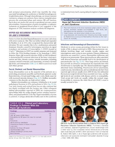
Facial, Skeletal, and Dental Abnormalities cryptococcosis, histoplasmosis, and coccidioidomycosis have been
Facial abnormalities seen in the majority of the patients are a reported. IgE levels are usually above 2000 IU/mL, but substantial
protruding, prominent mandible and forehead, apparent ocular fluctuations in IgE levels have been recorded over time, and the
hypertelorism, a broad nasal bridge, and a wide, fleshy nasal tip IgE levels do not correlate with disease activity or eosinophilia.
with increased interalar distance (Fig. 22.9). Midline anomalies Total serum IgG levels are usually within the normal range.
common in this disorder are high-arched palate. Eosinophilia is common; the white blood cell (WBC) count is
Skeletal abnormalities are common. Grimbacher et al. noted usually normal to low.
pathological fractures in 57% and scoliosis in 76% (Fig. 22.10).
Low bone density and cortical bone loss are also common but
not clearly correlated with the fracture rate. Other infrequent
skeletal abnormalities reported in HIES are craniosynostosis,
spina bifida, bifid rib, wedge-shaped lumbar vertebra, hemiver-
tebra, and pseudoarthritis of the hip. Hyperextensibility of joints
is common. A unique dental abnormality seen in this syndrome
TABLE 22.4 Clinical and Laboratory A B C D
Findings in Patients with the
Hyper-IgE Syndrome
Findings Incidence (%)
Eczema 100
High IgE levels (>2000 IU/mL) 97
Eosinophilia (>2 SD above the mean for normals) 93
Boils 87
Pneumonia 87 E F G H
Mucocutaneous candidiasis 83 FIG 22.9 Facial Abnormalities Seen in Patients With Hyper-IgE
Characteristic facies (in those ≥16 years) 83
Lung cysts 77 Recurrent Infection Syndrome (HIES). Prominent mandible
Scoliosis (for those ≥16 years) 76 and forehead, apparent hypertelorism, broad nasal bridge with
Hyperextensible joints 68 a wide nasal tip, and increased interalar distance are commonly
Delayed shedding of primary teeth 72 seen facial features of HIES. (With permission from Grimbacher
Bone fractures 57 B, Holland SM, Gallin JI, et al. Hyper-IgE syndrome with recurrent
infections—an autosomal dominant multisystem disorder. N Engl
Adapted from Grimbacher B, Holland SM, Gallin JI, et al. Hyper-IgE syndrome
disorder. N Engl J Med 1999; 340: 692. J Med 1999; 340: 692.)