
Page 356 - Clinical Immunology_ Principles and Practice ( PDFDrive )
P. 356
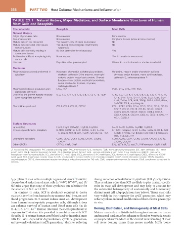
336          ParT TwO  Host Defense Mechanisms and Inflammation


         TABLE 23.1  Natural History, Major Mediators, and Surface Membrane Structures of Human
         Mast Cells and Basophils
          Characteristic              Basophils                            Mast Cells
          Natural History
          Origin of precursor cells   Bone marrow                          Bone marrow
          Site of maturation          Bone marrow                          Peripheral tissues (a few in bone marrow)
          Mature cells in the circulation  Yes (usually <1% of blood leukocytes)  No
          Mature cells recruited into tissues   Yes (during immunological, inflammatory   No
           from circulation             responses)
          Mature cells normally residing in   No (not detectable by microscopy)  Yes
           connective tissues
          Proliferative ability of morphologically   None reported         Yes (in certain circumstances)
           mature cells
          Life span                   Days (like other granulocytes)       Weeks to months (based on studies in rodents)

          Mediators
          Major mediators stored preformed in   Histamine, chondroitin sulfates-glucuronidase,   Histamine, heparin and/or chondroitin sulfates,
           cytoplasm                    elastase, cathepsin G-like enzyme, eosinophil   chymase and/or tryptase, many acid hydrolases,
                                        cationic protein, major basic protein, Charcot-  cathepsin G, carboxypeptidase A
                                        Leyden crystal protein, eosinophil peroxidase,
                                        some positive for tryptase, chymase,
                                        carboxypeptidase A
          Major lipid mediators produced upon   LTC 4 , PAF                PGD 2 , LTC 4 , LTB 4 , PAF, TXA 2
           appropriate activation
          Cytokines and growth factors released   IL-2, IL-3, IL-4, IL-5, IL-6, IL-9, IL-13, IL-15, TSLP  IL-1β, IL-2, IL-3, IL-4, IL-5, IL-6, IL-8, IL-9, IL-10, IL-11,
           upon appropriate activation                                      IL-13, IL-15, IL-16, IL-17A, IL-18, IL-22, IL-24, IL-25,
                                                                            IL-33, TNF-α, SCF, NGF, TGF-β, FGF-2, VEGF, IFN-α,
                                                                            GM-CSF, TSLP, amphiregulin
          Chemokines produced         CCL3, CCL4, CCL12, CXCL2             CCL1, CCL2, CCL3, CCL4, CCL5, CCL7, CCL8, CCL13,
                                                                            CCL17, CCL18, CCL19, CCL20, CCL22, CCL23,
                                                                            CCL25, CCL28, CXCL2, CXCL3, CXCL4, CXCL5,
                                                                            CXCL7, CXCL8, CXCL10, CXCL14, CXCL16, CXCL17,
                                                                            XCL1, CX3CL1
          Surface Structures
          Ig receptors                FcεRI, FcγRII (CDw32), FcγRIIB, FcγRIIIA  FcεRI, FcγRI, FcγRIIA, FcγRIIB, FcγRIIIA
          Cytokine/growth factor receptors  IL-1RIIb (CD121b), IL-2R (CD25), IL-3R, IL-4Rα,   KIT (SCF receptor), IL-3R, IL-4Rα, IL5Rα, IL-6R, IL-18R,
                                        IL-5Rα, IL-18R, IL-33R, TSLPR, GM-CSFRα, TrkA  IL-33R, IFN-γRα, TGF-β type-I and type-II β receptors,
                                                                            TrkA, T1/ST2/IL-1R4
          Chemokine receptors         CCR1, CCR2, CCR3, CCR5, CXCR1, CXCR2,   CCR1, CCR3, CCR4, CCR5, CXCR1, CXCR2, CXCR3,
                                        CXCR4                               CXCR4, CX3CR1
          Other GPCRs                 CRTH2, C3aR, C5aR                    EP3, BLT1, BLT2, cysLT1, PAF receptor, C3aR, C5aR
        LT, leukotriene; PG, prostaglandin; PAF, platelet-activating factor; TXA 2, thromboxane A 2; IL, interleukin; TSLP, thymic stromal lymphopoietin; SCF, stem cell factor; NGF, nerve
        growth factor; TGF-β, transforming growth factor-β; FGF-2, fibroblast growth factor-2; VEGF, vascular endothelial growth factor; IFN-α, interferon-α; GM-CSF, granulocyte
        macrophage–colony-stimulating factor; CCL, chemokine (C-C motif) ligand; CXCL, chemokine (C-X-C motif) ligand; XCL, chemokine (C motif) ligand; CX3CL, chemokine (C-X3-C
        motif) ligand; TrkA, tropomyosin receptor kinase A; CCR, C-C chemokine receptor; CXCR, C-X-C chemokine receptor; CX3CR, C-X3-C chemokine receptor; GPCRs, G protein–
        coupled receptors; CRTH2, chemoattractant receptor-homologous molecule expressed on Th2 cells; C3aR, complement component 3a receptor; C5aR, complement component 5a
        receptor.


                                                  9
        hyperplasia of mast cells in multiple organs and tissues.  However,   strong induction of leukotriene C 4  synthase (LTC 4 S) expression.
                                            v
                                                sh
                                                   sh
        the profound reduction of mast cells in W/W , W /W , and Sl/  Thus cytokines other than SCF are likely to play context-specific
        Sld mice argue that none of these cytokines can substitute for   roles in mast cell development and may help to account for
        the absence of SCF or CD117.                           the substantial heterogeneity of anatomically and functionally
           In contrast to mice, SCF is absolutely required to derive   distinct mast cell subpopulations (see below). Tissue mast cells
        mast cells in vitro from human bone marrow, fetal liver, or cord   vary widely in their capacity for cysLT generation, which may
        blood progenitors. IL-3 cannot induce mast cell development   reflect cytokine-induced modification of their effector phenotype
        from human hematopoietic progenitor cells, although it does   in vivo.
        can enhance survival of human cord blood mast cells (as do
                        10
        IL-4, IL-5, or IL-6).  Human intestinal mast cells proliferate in   Homing, Distribution, and Heterogeneity of Mast Cells
        response to IL-4 when it is provided in combination with SCF.   Mature mast cells are distributed throughout connective tissues
        Notably, IL-4 primes human cord blood and/or intestinal mast   and mucosal surfaces, often adjacent to blood or lymphatic vessels
        cells for FcεRI-dependent degranulation, cytokine generation,   or peripheral nerves. Much of the current understanding of mast
                                           11
        and cysteinyl leukotriene (cysLT) generation,  the latter reflecting   cell tissue homing comes from mouse models. MCPs home