
Page 360 - Clinical Immunology_ Principles and Practice ( PDFDrive )
P. 360
340 ParT TwO Host Defense Mechanisms and Inflammation
A
B
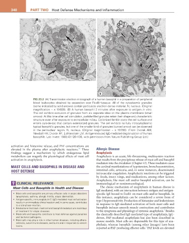
FIG 23.2 (A) Transmission electron micrograph of a human basophil in a preparation of peripheral
blood leukocytes obtained by separation over Ficoll-Hypaque. All of the cytoplasmic granules
(some indicated by solid arrows) contain particulate electron-dense material. N, nucleus. (Original
magnification ~ × 19 800). (B) A human basophil 2 minutes after exposure to antigen in vitro.
The cell exhibits extrusion of granules from six separate sites on the plasma membrane (small
arrows). At this time after cell stimulation, particle-filled granules retain their shape and characteristic
structure even after exposure to extracellular milieu. Cationized ferritin coats the cell surface and
enters culs-de-sac that contain exteriorized granules. The cell exhibits no fully intracytoplasmic
typical basophilic granules, but one of the smaller kind of granules (curved arrow) can be observed
in the perinuclear region. N, nucleus. (Original magnification ~ × 19 200). (From Dvorak AM,
Newball HH, Dvorak HF, Lichtenstein LM. Antigen-induced IgE-mediated degranulation of human
basophils. Lab Invest 1980;43:126–139, with permission from Nature Publishing Group Ltd.)
activation and histamine release, and PAF concentrations are
33
elevated in the plasma after anaphylactic reactions. These Allergic Disease
findings suggest a mechanism by which endogenous lipid Anaphylaxis
metabolism can magnify the physiological effects of mast cell Anaphylaxis is an acute, life-threatening, multisystem reaction
activation in anaphylaxis. that results from the precipitous release of mast cell and basophil
mediators into the circulation (Chapter 42). These mediators cause
MAST CELLS AND BASOPHILS IN DISEASE AND the cardinal manifestations of hypotension, bronchoconstriction,
HOST DEFENSE intestinal colic, urticaria, and, in some instances, disseminated
intravascular coagulation. Anaphylactic reactions can be triggered
by foods, insect stings, and medications, among other factors.
Anaphylaxis, like mast cell and/or basophil activation, can be
CLINICaL rELEVaNCE immunological or nonimmunological.
Mast Cells and Basophils in Health and Disease The classic mechanism of anaphylaxis in human disease is
IgE mediated, with an interaction between antigen and antigen-
• Mast cells and basophils are primary effector cells in atopic disorders specific IgE bound to FcεR1 on mast cells and/or basophils. In
(anaphylaxis, asthma, allergic rhinitis, atopic dermatitis). this instance, anaphylaxis can be considered a severe form of
• Antigen-specific, immunoglobulin E (IgE)-mediated mast cell activation type I hypersensitivity. Production of histamine and leukotrienes
results in an immediate clinical reaction and, in some cases, contributes
to a late-phase reaction. in response to IgE-mediated activation of both mast cells and
• Medications that block mast cell and basophil mediators are mainstays basophils induces smooth muscle contraction and contributes
3
of treatment for atopic diseases. to the symptoms and physiology of systemic anaphylaxis. Besides
• Mast cells and basophils contribute to host defense against parasites the classically described IgE-mediated type of anaphylaxis, IgG-
and bacterial pathogens. driven, PAF-mediated anaphylaxis has also been described in
• Mast cells may play a role in other human diseases, including athero- mouse models. Mast cells are dispensable for this type of ana-
sclerosis, autoimmune diseases, and burns and in responses to venom
toxins. phylaxis, whereas basophils (among other lineages) have been
3
reported as PAF-producing effector cells. PAF levels are elevated