
Page 361 - Clinical Immunology_ Principles and Practice ( PDFDrive )
P. 361
CHaPTEr 23 Mast Cells, Basophils, and Mastocytosis 341
B
A
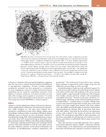
FIG 23.3 (A) Mast cell purified from human lung. The cell contains many cytoplasmic granules
with scroll-like substructural elements (solid arrows) and eight large non–membrane-bound lipid
bodies (open arrows). The plasma membrane has prominent folds. N, nucleus. (Original magnification
~ × 12 900). (B) An isolated human lung mast cell 10 minutes after exposure to anti-IgE in vitro.
Some degranulation channels (C), formed by fusion of membranes surrounding individual cytoplasmic
granules, contain altered granule matrix; others (EC) are empty. Cationized ferritin stains the
plasma membrane and the membranes of some empty degranulation channels (EC). The membranes
lining other channels (C) are unstained. A few unaltered scroll-containing granules (solid arrows)
remain. Numerous lipid bodies are also present (open arrows). The cytoplasm contains prominent
filaments. N, nucleus. (Original magnification ~ × 9 100). (From Galli SJ, Dvorak AM, Dvorak HF.
Prog Allergy 1984;34:1, with permission from Ann M. Dvorak.)
19
in the serum of patients with severe peanut anaphylaxis, suggesting prostanoids. The involvement of mast cells in early- and late-
that PAF may serve to amplify the physiological manifestations phase responses is supported by earlier studies using mast cell
of IgE-dependent anaphylaxis. Immune complex–mediated stabilizing cromone drugs, as well as anti-IgE.
complement activation has been proposed as a mechanism Immunohistological studies provide additional support for
for some anaphylactic reactions to drugs. Nonimmunological the role of mast cells in asthma. Bronchial biopsies from subjects
anaphylaxis can be caused by complement activation in the with asthma exhibit mast cells localizing to the airway smooth
absence of immune complexes or by direct activation of mast muscle bundles, a region generally lacking mast cells in biopsies
34
cells, often by drugs, such as opiates or vancomycin. As noted from healthy controls and from subjects with nonasthmatic
19
above, some of these (particularly muscle relaxants) may result eosinophilic bronchitis. Smooth muscle mast cell density cor-
from drugs that activate MRGPRX2 or related receptors. 32 related strongly with disease severity and with hyperresponsiveness
35
to methacholine, a cardinal feature of asthma. Notably, these
Asthma increases were observed irrespectively of atopic status of the study
Asthma is a chronic inflammatory disease of the airways character- subjects. In biopsy samples from subjects with severe asthma,
ized by variable airflow obstruction and airway hyperresponsive- mast cells exhibit substantially higher proportions with positive
ness (AHR) (Chapter 41). The most direct evidence for a immunostaining for chymase than in specimens from those
mechanistic contribution of mast cells to allergic asthma comes with mild asthma. Thus both the distribution and phenotype
from allergen challenge studies. In sensitized individuals, allergen of mast cells is altered in the airways of patients with severe
challenge causes an increase in mast cell granule contents in asthma. The mechanisms responsible for these alterations, and
bronchoalveolar lavage (BAL) fluid, including histamine, tryptase, the factors responsible for driving mast cell activation in severe
and cysLTs. About 60% of patients having an immediate, early- disease, particularly in individuals without atopy remain to be
phase asthmatic response will have a late-phase reaction beginning determined.
3–5 hours later, peaking around 8 hours, and resolving in about Mast cells may play an especially important role in aspirin-
24 hours. Mast cells contribute to both the early-phase and the exacerbated respiratory disease (AERD), a distinctive disorder
late-phase asthmatic response, via the release of histamine and in which asthma, severe rhinosinusitis, and nasal polyposis are
production of proinflammatory cytokines, leukotrienes, and associated with idiosyncratic respiratory reactions to aspirin and