
Page 364 - Clinical Immunology_ Principles and Practice ( PDFDrive )
P. 364
344 ParT TwO Host Defense Mechanisms and Inflammation
A B
C D
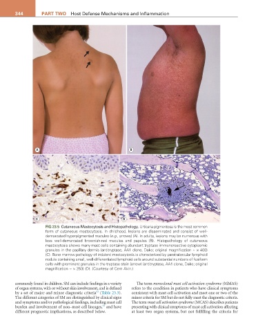
FIG 23.5 Cutaneous Mastocytosis and Histopathology. Urticaria pigmentosa is the most common
form of cutaneous mastocytosis. In childhood, lesions are disseminated and consist of well-
demarcated hyperpigmented macules (e.g., arrows) (A). In adults, lesions may be numerous with
less well-demarcated brownish-red macules and papules (B). Histopathology of cutaneous
mastocytosis shows many mast cells containing abundant tryptase immunoreactive cytoplasmic
granules in the papillary dermis (antitryptase, AA1 clone, Dako; original magnification ~ × 400)
(C). Bone marrow pathology of indolent mastocytosis is characterized by paratrabecular lymphoid
nodule containing small, well-differentiated lymphoid cells around substantial numbers of fusiform
cells with prominent granules in the tryptase stain (arrow) (antitryptase, AA1 clone, Dako; original
magnification ~ × 250) (D). (Courtesy of Cem Akin.)
commonly found in children. SM can include findings in a variety The term monoclonal mast cell activation syndrome (MMAS)
of organ systems, with or without skin involvement, and is defined refers to the condition in patients who have clinical symptoms
53
by a set of major and minor diagnostic criteria (Table 23.3). consistent with mast cell activation and meet one or two of the
The different categories of SM are distinguished by clinical signs minor criteria for SM but do not fully meet the diagnostic criteria.
and symptoms and/or pathological findings, including mast cell The term mast cell activation syndrome (MCAS) describes patients
52
burden and involvement of non–mast cell lineages, and have presenting with clinical symptoms of mast cell activation affecting
different prognostic implications, as described below. at least two organ systems, but not fulfilling the criteria for