
Page 363 - Clinical Immunology_ Principles and Practice ( PDFDrive )
P. 363
CHaPTEr 23 Mast Cells, Basophils, and Mastocytosis 343
Mastocytosis can be limited to skin or have widespread systemic Skin findings may be seen in both CM and SM, and the most
effects. Mast cell disorders cause a broad range of symptoms common skin-related symptom in patients with mastocytosis is
that can mimic a variety of other conditions, often making pruritus. Accumulations of mast cells in skin can present as a
diagnosis a challenge. variety of lesions. Urticaria pigmentosa (UP), also called macu-
lopapular cutaneous mastocytosis (MPCM), is the most common
Epidemiology skin manifestation of mastocytosis in both adults and children.
Mastocytosis can occur at any age, although it tends to have a Lesions of UP appear as small, tan to reddish-brown macules
more benign and transient course in children and often is limited or slightly elevated papules (Fig. 23.5A–C). UP is present more
to cutaneous manifestations. In a Polish case series of 100 children commonly in the less aggressive forms of SM.
with cutaneous mastocytosis (CM), 73% had onset of the disease Diffuse cutaneous mastocytosis (DCM) is characterized by
45
by age 6 months and 94% within the first year of life. Most edema and increased skin thickness, with or without a yellowish-
cases of systemic mastocytosis (SM) are diagnosed in middle brown coloration. DCM is rare, accounting for 1–3% of CM.
age. The exact prevalence of mastocytosis is unknown, but Cutaneous mastocytomas are well-demarcated flat or slightly
an estimate from a recent Danish population-based study is elevated lesions that may have a yellow or red-brown coloration.
46
approximately 1 : 10 000. With rare exceptions, mastocytosis Mastocytomas can be solitary or multiple and are typically 2–5 cm
does not appear to be inherited. in size. The least frequent form of CM (<1% of cases) is telan-
giectasia macularis eruptive perstans (TMEP). TMEP typically
Pathogenesis presents in adulthood and consists of tan/brown macules with
The manifestations of mastocytosis can result from mast cell telangiectasias.
mediator release (both chronic and episodic) and from excessive Lesions of UP, DCM, and cutaneous mastocytomas display
accumulation of mast cells in one or more tissues. Although the the Darier sign: localized urtication and redness of lesions fol-
molecular pathogenesis is incompletely understood, mastocytosis lowing rubbing, scratching, or stroking of skin. Mastocytomas
is frequently associated with somatic gain-of-function mutations can also precipitate severe systemic symptoms when rubbed. In
in KIT (CD117). The most common of these mutations is both CM and SM, mast cell mediator release can occur either
47
Asp816Val, or D816V. KIT-activating mutations lead to SCF- chronically or episodically, resulting in a variety of symptoms,
independent activation. Mast cells are thought to accumulate in including flushing, pruritus, shortness of breath, nausea, vomiting,
tissues because of clonal expansion and apoptotic defects of abdominal pain, diarrhea, hypotension, syncope, fatigue, and
KIT-mutated mast cells in mastocytosis. 6 headache. Anaphylaxis can be seen in both CM and SM. Interest-
ingly, urticaria and angioedema are uncommon in mastocytosis.
Clinical Features Gastrointestinal (GI) symptoms are particularly common in
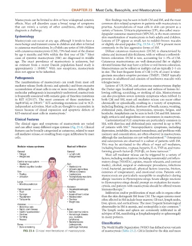
The clinical signs and symptoms of mastocytosis are varied SM, with diarrhea and abdominal pain reported in up to 80%
48
and can affect many different organ systems (Fig. 23.4). Clinical of patients with SM. Neuropsychiatric symptoms, including
features can be broadly categorized as cutaneous, related to mast depression, irritability, increased somnolence, and problems with
cell mediator release, or resulting from organ infiltration by mast memory and concentration, are often observed in mastocytosis,
49
cells. although the mechanisms are not well-understood. Osteopenia
and osteoporosis are observed in a subset of patients with SM.
This may be attributed to the effects of mast cell mediators,
Mediator release syndrome Mast cell infiltration
including histamine, tryptase, heparin, IL-6, TNF-α, and trans-
General General forming growth factor-β (TGF-β), on bone turnover. 50
• Fatigue • Lymph node Mast cell mediator release can be triggered by a variety of
• Weight loss enlargement
factors, including medications (including nonsteroidal antiinflam-
Central nervous system matory drugs [NSAIDs], opiates, muscle relaxants, and contrast
• Headache Skin media), alcohol, surgical or endoscopic procedures, infections
• Altered cognitive function • Mastocytoma
• Urticaria pigmentosa (viral, bacterial, parasitical), physical factors (exercise, friction,
Skin • Diffuse cutaneous extremes of temperature), and emotional stress. Patients with
• Pruritus mastocytosis mastocytosis are particularly susceptible to anaphylaxis during
• Urtication • Telangiectasia macularis
eruptiva perstans allergic reactions to Hymenoptera stings. Severe allergic reactions
Lungs to Hymenoptera stings should prompt an evaluation for masto-
• Bronchoconstriction
Abdomen cytosis, and patients with mastocytosis should be offered venom
Cardiovascular • Hepatosplenomegaly immunotherapy. 51
• Flush • Ascites Infiltration and/or proliferation of mast cells in organs other
• Syncope • Impaired liver function
• Hypotension • Malabsorption than the skin distinguish SM from CM. The organ systems most
• Tachycardia • Diarrhea often affected in SM include bone marrow, GI tract, lymph nodes,
• Weight loss
Abdomen liver, spleen, and cortical bone. The most frequent hematological
52
• Abdominal pain abnormality in SM is anemia, and eosinophilia is also common.
• Peptic ulcer disease Bones The lymph nodes and spleen are commonly infiltrated in all
• Gastric hypersecretion • Bone marrow lesions
• Diarrhea • Hematologic disease subtypes of SM, manifesting as lymphadenopathy or splenomegaly
• Vomiting (e.g., leukemia, in many patients.
• Nausea lymphoma)
• Skeletal lesions Classification
Bones (osteoporosis,
• Bone remodeling pathologic fractures) The World Health Organization (WHO) has defined seven variants
FIG 23.4 Clinical manifestations of mastocytosis. of mastocytosis (Table 23.2). CM is limited to the skin and more