
Page 493 - Clinical Immunology_ Principles and Practice ( PDFDrive )
P. 493
CHAPtER 34 Primary Antibody Deficiencies 473
Antigen dependent
M M
IgM
CVID CVID
XLA M G
Antigen independent
Mψ L M M G IgG
µ D HIGM CVID CVID
TdT E
XLA XLA M
µ 0
IGLL1 0 HIGM E IgE
Pro-B Pre-B Immature Mature CVID CVID
cell cell B cell B cell
HIGM M A
A IgA
IgAD IgAD
CVID CVID
Class Mature Plasma
switching B cell cell
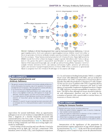
fIG 34.1 Defects in B-Cell Development Can Lead to Humoral Immune Deficiency. X-linked
agammaglobulinemia (XLA) and autosomal agammaglobulinemias (AGMs) result from either a
failure to generate a functional antibody receptor signaling complex or a failure to transmit signals
from this complex. These failures obstruct production of immature B cells. Hyper-IgM syndrome
(HIGM) results from either a failure to engage in proper cognate interactions with T cells or disrup-
tions in the genes that permit class-switch recombination or somatic hypermutation. These
failures inhibit class switching to IgG, IgA, and IgE, as well as limiting affinity maturation. Common
variable immune deficiency (CVID), IgG subclass deficiencies (IgSD), selective IgA deficiency
(IgAD), and transient hypogammaglobulinemia of infancy (THI) reflect a selective or generalized
failure to progress from the immature B-cell stage to the plasma cell stage.
KEY CoNCEPtS C3, C4, and mannose-binding lectin protein [MBL]), a complete
Hypogammaglobulinemia and blood count with differential (CBC/diff), and an erythrocyte
sedimentation rate (ESR). Lymphopenia is found most often in
Antibody Deficiency disorders that affect the production or function of T cells (Chapter
35) but can also occur in patients with CVID. Congenital absence
• The genetic mutations that underlie most primary antibody deficiencies
tend to affect genes that play key roles in the regulation of lymphocyte of an individual complement component will result in total
development and homeostasis. Clinically significant antibody deficiency absence of measurable complement-mediated hemolysis (Chapter
is not synonymous with hypogammaglobulinemia. 21). MBL deficiency increases susceptibility to respiratory infec-
3
• Serum immunoglobulin (Ig) concentrations vary widely with age. tions. The ESR is often, although not always, elevated in individu-
• Serum immunoglobulin levels vary with exposure to drugs (e.g., als with inflammatory disorders and can be useful in the
steroids), infectious agents, and other environmental stressors. evaluation of patients with a questionable or unclear history of
• A complete absence of IgG subclasses resulting from homozygous
deletions of heavy-chain genes has been observed in healthy recurrent or chronic infection.
individuals.
• Absence of a specific Vκ gene segment has been associated with
an increased risk of H. influenzae infection in spite of normal serum KEY CoNCEPtS
Ig levels. Testing for Immune Function
Testing for immune function should be performed:
• When the patient’s history suggests a rate or severity of infection
that exceeds normal expectations
expectations for normal individuals, when an opportunistic • When the organism responsible for infection is of low virulence or is
pathogen or one of a low virulence is responsible for an infection, considered to be an opportunistic pathogen
when a diagnosis of a disorder frequently associated with • When there is a diagnosis of a genetic syndrome or disorder associated
immunodeficiency has been made, or when there is a family with immune deficiency either in the patient or in the patient’s family
history of primary immunodeficiency. Table 34.2 illustrates four
levels of testing complexity and when such testing might com-
monly be undertaken. Interpretation of the significance of the quantitative Ig
Level I testing is both revealing and cost effective. It includes determinations requires appreciation of age-related changes in
4,5
measuring serum Ig (IgM, IgG, and IgA), complement (50% Ig concentrations (Fig. 34.2). At the end of the second trimester
hemolytic power of serum [CH 50 ] and complement components of pregnancy, active transport of IgG across the placental barrier