
Page 660 - Clinical Immunology_ Principles and Practice ( PDFDrive )
P. 660
634 Part five Allergic Diseases
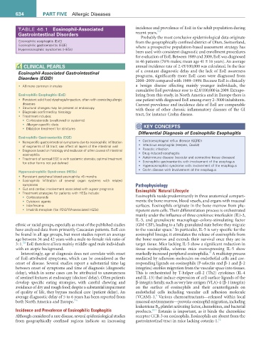
TABLE 46.1 eosinophil-associated incidence and prevalence of EoE in the adult population during
7-9
Gastrointestinal Disorders recent years.
Probably the most conclusive epidemiological data originate
Eosinophilic esophagitis (EoE) from the geographically confined district of Olten, Switzerland,
Eosinophilic gastroenteritis (EGE) where a prospective population-based assessment strategy has
Hypereosinophilic syndromes (HESs)
been used with consistent diagnostic and enrollment procedures
for evaluation of EoE. Between 1989 and 2009, EoE was diagnosed
in 46 patients (76% males; mean age 41 ± 16 years). An average
CLiNiCaL PearLS annual incidence rate of 2.45/100,000 was calculated. In the face
Eosinophil-Associated Gastrointestinal of a constant diagnostic delay and the lack of EoE awareness
Disorders (EGID) programs, significantly more EoE cases were diagnosed from
2000–2009 compared with 1989–1999. Because EoE is clinically
• All more common in males a benign disease affecting mainly younger individuals, the
cumulative EoE prevalence rose to 42.8/100,000 in 2009. Extrapo-
eosinophilic esophagitis (eoe) lating from this study, in North America and in Europe there is
• Persistent solid food dysphagia/impaction, often with coexisting allergic one patient with diagnosed EoE among every 2–3000 inhabitants.
diseases Current prevalence and incidence data of EoE are comparable
• Structural changes may be present at endoscopy with those of other chronic inflammatory diseases of the GI
• Diagnosis confirmed by histology tract, for instance Crohn disease.
• Treatment includes:
• Corticosteroids (swallowed or systemic)
• Allergen-specific diets KeY CONCePtS
• Dilatation treatment for strictures
Differential Diagnosis of Eosinophilic Esophagitis
eosinophilic Gastroenteritis (eGe)
• Nonspecific gastrointestinal symptoms due to eosinophilic infiltration • Gastroesophageal reflux disease (GERD)
of segments of GI tract; can affect all layers of the intestinal wall • Infectious esophagitis (Herpes, Candida)
• Diagnosis based on histology and exclusion of other causes of intestinal • Parasitic infection
eosinophilia • Drug-induced esophagitis
• Treatment of serosal EGE is with systemic steroids; optimal treatment • Autoimmune disease (vascular and connective tissue diseases)
for other forms not yet defined • Eosinophilic gastroenteritis with involvement of the esophagus
• Hypereosinophilic syndrome with involvement of the esophagus
• Crohn disease with involvement of the esophagus
Hypereosinophilic Syndromes (HeSs)
• Persistent peripheral blood eosinophilia >6 months
• Eosinophilic infiltration of several organ systems with related
symptoms Pathophysiology
• Gut and cardiac involvement associated with a poor prognosis Eosinophils’ Natural Lifecycle
• Treatment strategies for patients with HESs include:
• Corticosteroids Eosinophils reside predominantly in three anatomical compart-
• Cytotoxic agents ments: the bone marrow, blood vessels, and organs with mucosal
• Interferon-α surfaces. Eosinophils originate in the bone marrow from plu-
• Imatinib mesylate (for PDGFRA-associated HESs) ripotent stem cells. Their differentiation process is orchestrated
mainly under the influence of three cytokines: interleukin (IL)-3,
IL-5, and granulocyte macrophage–colony-stimulating factor
ethnic or racial groups, especially as most of the published studies (GM-CSF), leading to a fully granulated state before they migrate
1
have analyzed data from primarily Caucasian patients. EoE can to the vascular space. In particular, IL-5 is very specific for the
be found in all age groups, but most studies report an average eosinophil lineage; it stimulates the release of eosinophils from
age between 34 and 42 years with a male-to-female risk ratio of the bone marrow and extends their survival once they are in
7,8
3 : 1. EoE therefore affects mainly middle-aged male individuals target tissue. Mice lacking IL-5 show a significant reduction in
with an atopic background. tissue eosinophilia, whereas mice overexpressing IL-5 show
10
Interestingly, age at diagnosis does not correlate with onset markedly increased peripheral eosinophilia. A multistep process
of EoE-attributed symptoms, which can be considered as the mediated by adhesion molecules on endothelial cells and cor-
onset of disease. Several studies report a substantial time lag responding ligands on eosinophils (P-selectin and β-1 and β-2
between onset of symptoms and time of diagnosis (diagnostic integrins) enables migration from the vascular space into tissues.
delay), which in some cases can be attributed to unawareness This is orchestrated by T-helper cell-2 (Th2) cytokines (IL-4
of sentinel features at endoscopy (doctors’ delay). Often patients and IL-13) that induce expression of cell surface ligands of the
develop specific eating strategies, with careful chewing and β-integrin family, such as very late antigen (VLA)-4 (β-1 integrin)
avoidance of dry and rough food; despite a substantial impairment on the surface of eosinophils and their counterligands on
of quality of life, they bypass medical care (patient delay). An endothelial cells including vascular cell adhesion molecule
9
average diagnostic delay of 5 to 6 years has been reported from (VCAM)-1. Various chemoattractants—released within local
both North America and Europe. 7-9 mucosal environments—provoke eosinophil migration, including
leukotriene B 4 , platelet activating factor, chemokines, and bacterial
Incidence and Prevalence of Eosinophilic Esophagitis products. 9,11 Eotaxin is important, as it binds the chemokine
Although considered a rare disease, several epidemiological studies receptor CCR-3 on eosinophils. Eosinophils are absent from the
from geographically confined regions indicate an increasing gastrointestinal tract in mice lacking eotaxin-1. 12