
Page 661 - Clinical Immunology_ Principles and Practice ( PDFDrive )
P. 661
CHaPter 46 Eosinophil-Associated Gastrointestinal Disorders 635
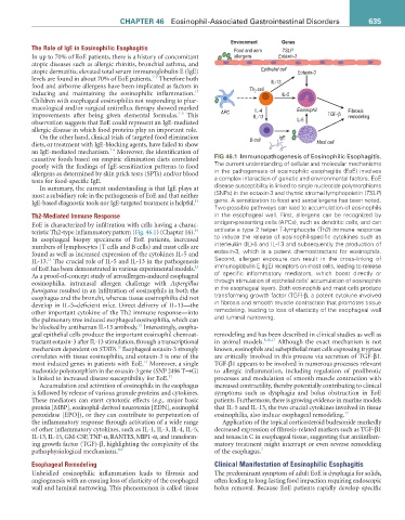
Environment Genes
The Role of IgE in Eosinophilic Esophagitis Food and aero TSLP
In up to 70% of EoE patients, there is a history of concomitant allergens Eotaxin-3
atopic diseases such as allergic rhinitis, bronchial asthma, and
atopic dermatitis; elevated total serum immunoglobulin E (IgE) Epithelial cell Eotaxin-3
7-9
levels are found in about 70% of EoE patients. Therefore both IL-13
food and airborne allergens have been implicated as factors in Th 2 cell
13
inducing and maintaining the eosinophilic inflammation. IL-5
Children with esophageal eosinophilia not responding to phar-
macological and/or surgical antireflux therapy showed marked IL-4 Eosinophil Fibrosis
7-9
improvements after being given elemental formulas. This APC IL-13 TGF-β remodeling
observation suggests that EoE could represent an IgE-mediated IL-5
allergic disease in which food proteins play an important role.
On the other hand, clinical trials of targeted food elimination B cell IgE
diets, or treatment with IgE-blocking agents, have failed to show Mast cell
7-9
an IgE-mediated mechanism. Moreover, the identification of
causative foods based on empiric elimination diets correlated fiG 46.1 Immunopathogenesis of Eosinophilic Esophagitis.
poorly with the findings of IgE-sensitization patterns to food The current understanding of cellular and molecular mechanisms
allergens as determined by skin prick tests (SPTs) and/or blood in the pathogenesis of eosinophilic esophagitis (EoE) involves
tests for food-specific IgE. a complex interaction of genetic and environmental factors. EoE
In summary, the current understanding is that IgE plays at disease susceptibility is linked to single nucleotide polymorphisms
most a subsidiary role in the pathogenesis of EoE and that neither (SNPs) in the eotaxin-3 and thymic stromal lymphopoietin (TSLP)
IgE-based diagnostic tools nor IgE-targeted treatment is helpful. 13 gene. A sensitization to food and aeroallergens has been noted.
Two possible pathways can lead to accumulation of eosinophils
Th2-Mediated Immune Response in the esophageal wall. First, allergens can be recognized by
EoE is characterized by infiltration with cells having a charac- antigen-presenting cells (APCs), such as dendritic cells, and can
14
teristic Th2-type inflammatory pattern (Fig. 46.1) (Chapter 16). activate a type 2 helper T-lymphocyte (Th2) immune response
In esophageal biopsy specimens of EoE patients, increased to induce the release of eosinophil-specific cytokines such as
numbers of lymphocytes (T cells and B cells) and mast cells are interleukin (IL)-5 and IL-13 and subsequently the production of
found as well as increased expression of the cytokines IL-5 and eotaxin-3, which is a potent chemoattractant for eosinophils.
14
IL-13. The crucial role of IL-5 and IL-13 in the pathogenesis Second, allergen exposure can result in the cross-linking of
15
of EoE has been demonstrated in various experimental models. immunoglobulin E (IgE) receptors on mast cells, leading to release
As a proof-of-concept study of aeroallergen-induced esophageal of specific inflammatory mediators, which boost directly or
eosinophilia, intranasal allergen challenge with Aspergillus through stimulation of epithelial cells’ accumulation of eosinophils
fumigatus resulted in an infiltration of eosinophils in both the in the esophageal layers. Both eosinophils and mast cells produce
esophagus and the bronchi, whereas tissue eosinophilia did not transforming growth factor (TGF)-β, a potent cytokine involved
develop in IL-5–deficient mice. Direct delivery of IL-13—the in fibrosis and smooth muscle contraction that promotes tissue
other important cytokine of the Th2 immune response—into remodeling, leading to loss of elasticity of the esophageal wall
the pulmonary tree induced esophageal eosinophilia, which can and luminal narrowing.
15
be blocked by antihuman IL-13 antibody. Interestingly, esopha-
geal epithelial cells produce the important eosinophil chemoat- remodeling and has been described in clinical studies as well as
tractant eotaxin-3 after IL-13 stimulation, through a transcriptional in animal models. 8,16,17 Although the exact mechanism is not
10
mechanism dependent on STAT6. Esophageal eotaxin-3 strongly known, eosinophils and subepithelial mast cells expressing tryptase
correlates with tissue eosinophilia, and eotaxin-3 is one of the are critically involved in this process via secretion of TGF-β1.
12
most induced genes in patients with EoE. Moreover, a single TGF-β1 appears to be involved in numerous processes relevant
nucleotide polymorphism in the eotaxin-3 gene (SNP 2496 T→G) to allergic inflammation, including regulation of profibrotic
is linked to increased disease susceptibility for EoE. 12 processes and modulation of smooth muscle contraction with
Accumulation and activation of eosinophils in the esophagus increased contractility, thereby potentially contributing to clinical
is followed by release of various granule proteins and cytokines. symptoms such as dysphagia and bolus obstruction in EoE
These mediators can exert cytotoxic effects (e.g., major basic patients. Furthermore, there is growing evidence in murine models
protein [MBP], eosinophil-derived neurotoxin [EDN], eosinophil that IL-5 and IL-13, the two crucial cytokines involved in tissue
peroxidase [EPO]), or they can contribute to perpetuation of eosinophilia, also induce esophageal remodeling. 17
the inflammatory response through activation of a wide range Application of the topical corticosteroid budesonide markedly
of other inflammatory cytokines, such as IL-1, IL-3, IL-4, IL-5, decreased expression of fibrosis-related markers such as TGF-β1
IL-13, IL-15, GM-CSF, TNF-α, RANTES, MIP1-α, and transform- and tenascin C in esophageal tissue, suggesting that antiinflam-
ing growth factor (TGF)-β, highlighting the complexity of the matory treatment might interrupt or even reverse remodeling
pathophysiological mechanisms. 8,9 of the esophagus. 7
Esophageal Remodeling Clinical Manifestation of Eosinophilic Esophagitis
Unbridled eosinophilic inflammation leads to fibrosis and The predominant symptom of adult EoE is dysphagia for solids,
angiogenesis with an ensuing loss of elasticity of the esophageal often leading to long-lasting food impaction requiring endoscopic
wall and luminal narrowing. This phenomenon is called tissue bolus removal. Because EoE patients rapidly develop specific